All posts by The Trauma Pro

Everything You Wanted To Know About: Cranial Bone Flaps

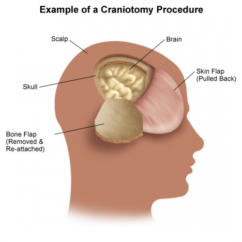
Patients with severe TBI frequently undergo surgical procedures to remove clot or decompress the brain. Most of the time, they undergo a craniotomy, in which a bone flap is raised temporarily and then replaced at the end of the procedure.

But in decompressive surgery, the bone flap cannot be replaced because doing so may increase intracranial pressure. What to do with it?

There are four options:

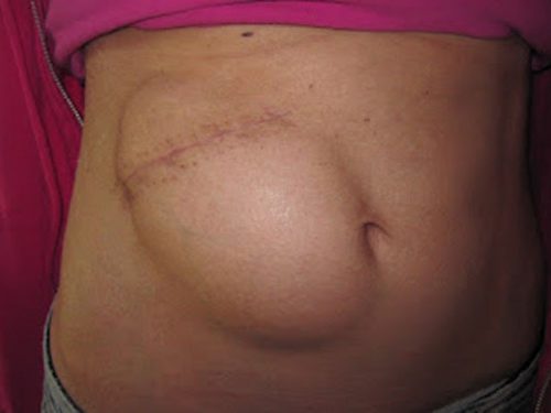
  1. The piece of bone can buried in the subcutaneous tissue of the abdominal wall. The advantage is that it can’t get lost. Cosmetically, it looks odd, but so does having a bone flap missing from the side of your head. And this technique can’t be used as easily if the patient has had prior abdominal surgery.

2. Some centers have buried the flap in the subgaleal tissues of the scalp on the opposite side of the skull. The few papers on this technique demonstrated a low infection rate. The advantage is that only one surgical field is necessary at the time the flap is replaced. However, the cosmetic disadvantage before the flap is replaced is much more pronounced.

3. Most commonly, the flap is frozen and “banked” for later replacement. There are reports of some mineral loss from the flap after replacement, and occasional infection. And occasionally the entire piece is misplaced. Another disadvantage is that if the patient moves away or presents to another hospital for flap replacement, the logistics of transferring a frozen piece of bone are very challenging.

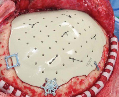
4. Some centers just throw the bone flap away. This necessitates replacing it with some other material like metal or plastic. This tends to be more complicated and expensive, since the replacement needs to be sculpted to fit the existing gap.

So which flap management technique is best? Unfortunately, we don’t know yet, and probably never will. Your neurosurgeons will have their favorite technique, and that will ultimately be the option of choice.

Reference: Bone flap management in neurosurgery. Rev Neuroscience 17(2):133-137, 2009.

The  March Trauma MedEd Newsletter Is Available!

This month’s newsletter contains a veritable potpourri of topics that I found interesting. I hope you do, as well. Here are the topics covered:

  • Prehospital lactate levels: useful or not?
  • Does aspirin for DVT prophylaxis work?
  • The nuts and bolts of Duplex ultrasound
  • An 11 year experience with endovascular repair of aortic injuries
  • Use of MRI after CT scanning of the cervical spine

Subscribers received this issue last week. Subscribe now and be sure to get the next issue early.  So sign up for early delivery now by clicking here!

Click here to download the current issue

Pick up back issues here!


Pet Peeve: “High Index of Suspicion”

How often have you heard this phrase in a talk or seen it in a print article:

“Maintain a high index of suspicion”

What does this mean??? It’s been popping up in papers and textbooks for at least the last 20 years. And to me, it’s meaningless. You try to figure out that sentence!

An index is a number, usually mathematically derived in some way. Yet whenever I see or hear this phrase, it doesn’t really apply to anything that is quantifiable. What the author is really referring to is “a high level of suspicion”, not an index.

This term has become a catch-all to caution the reader or listener to think about a (usually) less common diagnostic possibility. As trauma professionals, we are advised to do this about so many things, it really has become sad and meaningless.

Bottom line: Don’t use this phrase in your presentations or your writing. It’s stupid. And feel free to chide any of your colleagues who do. Give us some concrete tips so we don’t miss the diagnosis!

Reference: High index of suspicion. Ann Thoracic Surg 64:291-292, 1997.

Are State Trauma Systems Cost-Effective?

Every state in the US now has a formal trauma system. Several studies are available that document the advantages of these systems in terms of outcomes and survival. Trauma professionals get this. But the governmental agencies and legislators who help create, fund, and maintain them tend to focus on cost as well.

Arkansas was the last state in the union to implement a trauma system. A study in press from the University of Arkansas details their experience. They examined the impact of the new system on mortality, patient care, and attempted to calculate a return on investment from the taxpayers in an effort to show the added value.

The study was commissioned by the Arkansas Department of Health and carried out by the state Trauma Advisory Council. It was led by out of state investigators in an effort to maintain impartiality. A comprehensive review of records was performed by a panel of 5 surgeons, 1 emergency physician, 2 trauma program managers, 1 ground and 1 flight paramedic. Preventable and potentially preventable deaths were identified and analyzed in depth. Value of life lost was calculated by using a conservative $100,000 per year lost. A total of 290 charts were reviewed pre-system, and 382 post-trauma system implementation using proportional sampling of about 2500 trauma deaths in one year.

Here are the factoids:

  • A significantly higher percentage of patients were triaged to Level I trauma centers after the system was implemented
  • Preventable mortality was decreased from 30% to 14% (!!)
  • This means that 79 extra lives were saved due to implementation of the trauma system
  • Non-preventable deaths with opportunity for improvement remained constant at about 55%
  • Non-preventable deaths without opportunity for improvement increased from 16% to 38% (!)
  • Using the most conservative VLL calculation, this equates to $2.4M in savings per life saved
  • This adds up to $186M in savings to the taxpayers of Arkansas, a 9-fold return on their investment of $20M in the trauma system. 

Bottom line: Wow! This nicely done studies gives us excellent insight into the hows and whys of the value of an organized state trauma system. It is likely that the triage system directed more patients to the most appropriate level of care, leading to fewer preventable deaths. And it enticed hospitals to up their game and make the move toward formal trauma center designation. This improved education and training at those centers, leading to better patient care.

There is a wealth of information in this study, and I recommend that everyone with an interest in or are already participating in their state trauma system read it in its entirety. Hospitals that are reluctant to join or are lagging in meeting criteria need to recognize that they are not serving their communities as well as they think. And legislators must realize that the financial impact of even a small investment has real and significant consequences to their constituents.

Related posts:

Reference: Does the Institution of a Statewide Trauma
System Reduce Preventable Mortality and Yield
a Positive Return on Investment for Taxpayers? JACS in press, 2017.

Retained Hemothorax Part 4: The Practice Guideline

Over the last three days, I reviewed some data on lytics at the request of some of my readers. Then I looked at a paper describing one institution’s experience dealing with retained hemothorax, including the use of VATS. But there really isn’t much out there on how to roll all this together.

Until now. The trauma group at Vanderbilt has a paper in press describing their experience with a home-grown practice guideline for managing retained hemothorax.  Here’s what it looks like:

I know it’s small, so just click it to download a pdf copy. I’ve simplified the flow a little as well.

All stable patients with hemothorax admitted to the trauma service were included over a 2.5 year period. The practice guideline was implemented midway through this study period. Before implementation, patients were treated at the discretion of the surgeon. Afterwards, the practice guideline was followed.

Here are the factoids:

  • There were an equal number of patients pre- and post-guideline implementation (326 vs 316)
  • An equal proportion of each group required an initial intervention, generally a chest tube (69% vs 65%)
  • The number of patients requiring an additional intervention (chest tube, VATS, lytics, etc) decreased significantly from 15% to 9%
  • Empyema rate was unchanged at 2.5%
  • Use of VATS decreased significantly from 8% to 3%
  • Use of catheter guided drainage increased significantly from 0.6% to 3%
  • Hospital length of stay was the same, ranging from 4 to 11 days (much shorter than the lytics studies!)

Bottom line: This is how design of practice guidelines is supposed to work. Identify a problem, typically a clinical issue with a large amount of provider care variability. Look at the literature. In general, find it of little help. Design a practical guideline that covers the major issues. Implement, monitor, and analyze. Tweak as necessary based on lessons learned. If you wait for the definitive study to guide you, you’ll be waiting for a long time.

This study did not significantly change outcomes like hospital stay or complications. But it did decrease the number of more invasive procedures and decreased variability of care, with the attendant benefits from both of these. It also dictates more selective (and intelligent) use of additional tubes, catheters, and lytics. 

I like this so much that I plan to adopt it at my center!

Download the practice guideline here.

Related posts:

Posts in this series:

Reference: Use of an evidence-based algorithm for patients with traumatic hemothorax reduces need for additional interventions. J Trauma, in press, December 14, 2016.