All posts by The Trauma Pro

IV Contrast and Trauma – Revisited

We use CT scanning in trauma care so much that we tend to take it (and its safety) for granted. I’ve written quite a bit about thoughtful use of radiographic studies to achieve a reasonable patient exposure to xrays. But another thing to think about is the use of IV contrast.

IV contrast is a hyperosmolar solution that contains some substance (usually an iodine compound) that is radiopaque to some degree. It has been shown to have a significant impact on short-term kidney function and in some cases can cause renal failure.

Here are some facts you need to know:

  • Contrast nephrotoxicity is defined as a 25% increase in serum creatinine, usually within the first 3 days after administration
  • There is usually normal urine output and minimal to no proteinuria
  • In most cases, renal function returns to normal after 3-4 days
  • Nephrotoxicity almost never occurs in people with normal baseline kidney function
  • Large or repeated doses given within 72 hours greatly increase risk for toxicity
  • Old age and pre-existing diabetic renal impairment also greatly increase risk

If you must give contrast to a patient who is at risk, make sure they are volume expanded (tough in trauma patients), or consider giving acetylcysteine or using isosmolar contrast (controversial, may still cause toxicity).

Bottom line: If you are considering contrast CT, try to get a history to see if the patient is at risk for nephrotoxicity. Also consider all of the studies that will be needed and try to consolidate your contrast dosing. For example, you can get CT chest/abdomen/pelvis and CT angio of the neck with one contrast bolus. Consider low dose contrast injection if the patient needs formal angiographic studies in the IR suite. And finally, consider what changes will be made if the study is positive. For example, if a CT angio of the neck for blunt carotid/vertebral injury is being considered, the intervention for a positive result is usually just aspirin. Since this is a very benign medication, why not forgo the scan and just start aspirin if there is a significant risk of kidney injury from the contrast. Always think about the global needs of your patient and plan accordingly (and safely).

Reference: Contrast media and the kidney. British J Radiol 76:513-518, 2003.

Button Batteries: Part 2 – Getting Them Out

In my last post, I detailed how to suspect and image a button battery ingestion. In this one, I’ll describe how to extract them, and how quickly it’s necessary.

When batteries come to rest and are surrounded by moist mucosal tissue, a current arc is generated around the two sides of the button. This releases heat, which coagulates the surrounding tissue. Depending on the location, closeness of contact, and the duration, these burn injuries may extend into underlying tissue. This is of particular significance in the esophagus, which is in close proximity to the thoracic aorta.

Here’s a simple demonstration you can do at home with some lunch meat:

Here are guidelines for what to do when you encounter pediatric patients who have ingested a button battery:

  • If the child is experiencing bleeding from the upper GI tract, activate your trauma team. The child may have an aorto-esophageal fistula. If there is no active bleeding, obtain a chest x-ray to assess the battery’s position. If there is active bleeding, proceed to the OR (preferably a hybrid room if you have one) and use fluoro to locate the battery. If bleeding persists, call appropriate pediatric surgical specialists (surgery, CV surgery, GI), activate your massive transfusion protocol, and consider tamponade with a Blakemore tube (remember those?) or a urinary catheter if you don’t have one.
  • No bleeding from the upper GI tract? If the battery is large (>20mm) and/or the child is small (<5 years), and is lodged in the esophagus, proceed immediately to OR and remove endoscopically.
  • Batteries in the stomach are of less concern. They will generally pass if <20mm. A repeat x-ray after 48 hours should be obtained for larger batteries. If still in the stomach, they should be removed endoscopically. Smaller batteries will usually pass, and should be re-imaged after two weeks to confirm this.

References:

  • Button battery and magnet ingestions in the pediatric patient.  Curr Opin Pediatrics 30:653-659, 2018.
  • Management of ingested foreign bodies in children: a clinical report of the NASPGHAN Endoscopy Committee. J Pediatric Gastroenterol Nutr 60:562-574, 2015.

Button Batteries: Part 1

I know what you are saying. Button batteries? Trauma? Not too many adult trauma professionals have seen or heard of this. But those who care for pediatric patients should be very familiar. If the importance of this seemingly minor problem is ignored, the results can be catastrophic.

Kids eat stuff, and not just food. The smaller ones always seem to be putting things in their mouths. Foreign body ingestion (or insertion into other orifices) is a common presentation at pediatric emergency departments. Unfortunately, the fact that a battery has been eaten may not be appreciated by the parents.  The child may be brought in with  nonspecific GI or respiratory symptoms.

As soon as a battery ingestion is known or suspected, a two-view chest x-ray is needed. This should show both chest and upper abdomen in order to visualize both esophagus and stomach. Separate chest and abdominal images may be required if the child is too large for a single shot. Two views (AP and lateral) are important because the nature of the foreign body may not be appreciated if the battery is seen edge-on.

If you are fortunate enough to image the battery “face-on”, you may see a telltale halo sign. Because of the way these batteries are put together, there are two metal sides that have a slight difference in overlap.

You’ve made the diagnosis! So now what? And how quickly? I’ll deal with this in my next post.

How Fast Can You Warm Up A Hypothermic Patient?

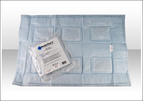
For most places, including Minnesota, hypothermia time is just about over! However, this trauma problem can occur nearly anywhere and at any time. And especially during a massive resuscitation. The optimal way to warm paitients has been debated for years. A number of very interesting techniques have been devised. Ever wonder how fast / effective they are?

I’ve culled data from a number of sources, and here is a summary what I found. And of course, the disclaimer: “your results may vary.”

Warming Technique Rate of Rewarming
Passive external (blankets, lights) 0.5° C / hr
Active external (lights, hot water bottle) 1 – 3° C / hr
Bair Hugger (a 3M product, made in Minnesota of course!) 2.4° C / hr
Hot inspired air in ET tube 1° C / hr
Fluid warmer 2 – 3° C / hr
GI tract irrigation (stomach or colon, 40° C fluid, instill for 10 minutes, then evacuate) 1.5 -3° C / hr
Peritoneal lavage (instill for 20-30 minutes) 1 – 3° C / hr
Thoracic lavage (2 chest tubes, continuous flow) 3° C / hr
Continuous veno-venous rewarming 3° C / hr
Continuous arterio-venous rewarming 4.5° C / hr
Mediastinal lavage (thoracotomy) 8° C / hr
Cardiopulmonary bypass 9° C / hr
Warm water immersion (Hubbard or therapy tank) 20° C / hr

In The Next Trauma MedEd Newsletter: Potpourri

The next issue of Trauma MedEd will be sent out to subscribers at the end of the week, and will cover a whole bunch of what I call Potpourri. These are some tidbits that I find fascinating and sometimes odd.

Topics will include:

  • Trauma and the gut microbiome
  • “Single look trauma laparotomy” (as opposed to damage control laparotomy) and postop complications
  • Pneumothorax: how big is too big?
  • The best prehospital stretchers for rapid extraction in MCIs
  • and more!

I’ve pushed the solid organ injury update issue out to next month. There’s a lot of stuff to cover, and quite a few changes have occurred over the years.

As always, this month’s issue will go to all of my subscribers first. If you are not yet one of them, click this link right away to sign up now and/or download back issues.

Unfortunately, non-subscribers will have to wait until I release the issue on this blog, in mid-April. So sign up now!