Help! My Consultant Won’t Come In To See A Patient!

Consultants provide very important services to trauma patients in the ED and inpatient settings. The trauma professionals managing those patients can’t know everything (although we sometimes think we do). But occasionally our patients present issues that require evaluation by other experts in order to guarantee excellent care.

Sometimes our consultants want to do too much, or make recommendations that are not really in their area of expertise (e.g. a cardiologist evaluating a cardiac contusion). See the related post link below for tips on this situation.

But sometimes you know what the patient needs, but the consultant doesn’t agree or doesn’t do what you expect. Or they don’t want to come in when called. What to do?

Here are some tips:

The patient is in the ED and the consultant won’t come in to see the patient.

  • Are they right? Does that problem really need to be dealt with in the ED in the middle of the night? Many simple fractures and wounds do not need immediate attention. They can be dressed/splinted, the patient reassured, and instructed to see the consultant in the clinic the next day.
  • Is your knowledge of current management of the condition correct? Perhaps it has evolved, and it is now commonplace to temporize and deal with the problem as an outpatient during business hours. Make sure you are up on the current literature.

The patient is in the ED and the consultant won’t come in to see the patient, and you are sure that they should! Now what?

  • Call them personally (not a resident, midlevel provider, or any other intermediary) and clearly and concisely explain the situation, and your assessment of why the problem needs their immediate attention.
  • Listen to or elicit their rationale for not seeing the patient. If legitimate, this may help educate you and modify your future management of similar patients. If the rationale is not legitimate, inform them (tactfully) that this is at odds with your education/training/experience with other providers. Ask them to further explain, if they can.

If they still won’t come in despite what you think is a legitimate need, then you must calculate a quick risk:benefit ratio. Will any patient harm occur if the consultant does not see the patient? And what is the professional damage that you will incur if you move on to the next steps. If you believe that harm will occur, here are your options, from least to most damaging to your professional status at the hospital:

  • Contact another consultant in the same or overlapping specialty (if there is one). Apologize for the fact that you know they are not on call, and explain the situation.
  • Appeal to a higher authority. Contact the trauma medical director, service chief, or hospital administrator and see if they can intervene.
  • Explain to the consultant that you truly believe that harm will occur, and you will have to document that fact in the medical record as well as their failure to respond. In some cases, this will shake them loose, but they will certainly be pissed.
  • If all else fails, see if you can find a service that will help you by accepting the patient as an admission so they can be managed appropriately the next day. But then follow through by reporting the event to appropriate people including chief of staff, chief medical officer, VPMA, hospital quality department, and risk management. This is the nuclear option, so be prepared for the fallout.

Bottom line: This is not a fun situation to find yourself in. Good luck!

New Technology: Using AI To Interpret Pelvic X-rays

Look out, radiologists! The computers are coming for you!

Radiologists use their extensive understanding of human anatomy and combine it with subtle findings they see on x-ray shadow pictures. In doing this, they can identify a wide variety of diseases, anomalies, and injuries. But as we have seen with vision systems and game playing (think chess), computers are getting pretty good at doing this as well.

Is it only a matter of time until computer artificial intelligence (AI) starts reading x-rays?  Look at how good they already are at interpreting EKGs. The trauma group at Stanford paired up with the Chang Gung Memorial Hospital in Taiwan to test the use of AI for interpreting images to identify a specific set of common pelvic fractures.

The Stanford group used a deep learning neural network (XCeption) to analyze source x-rays (standard A-P pelvis images) from Chang Gung. These x-rays were divided into training and testing cohorts. The authors also applied different degrees of blurring, brightness, rotation, and contrast adjustment to the training set in order to help the AI overcome these issues when interpreting novel images.

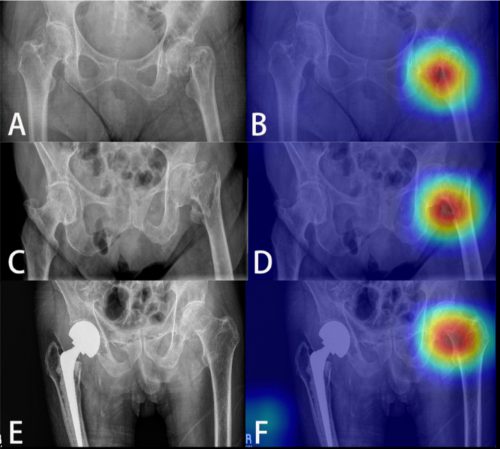
The AI interpreted the test images with a very high degree of sensitivity, specificity, accuracy, and predictive values, with all of them over 0.90. The algorithms generated a “heat map” that showed the areas that were suspicious for fracture. Here are some examples with the original x-ray on the left and the heat map on the right:

The top row shows a femoral neck fracture, the middle row an intertrochanteric fracture, and the bottom row another femoral neck fracture with a contralateral implant. All were handily identified by the AI.

AI applications are usually only as good as their training sets. In general, the bigger the better so they can gain a broader experience for more accurate interpretation. So it is possible that uncommon, subtle fractures could be missed. But remember, artificial intelligence is meant to supplement the radiologist, not replace him or her. You can all breathe more easily now.

This technology has the potential for broader use in radiographic interpretation. In my mind, the best way to use it is to first let the radiologist read the images as they usually do. Once they have done this, then turn on the heat map so they can see any additional anomalies the AI has found. They can then use this information to supplement the initial interpretation.

Expect to see more work like this in the future. I predict that, ultimately, the picture archiving and communications systems (PACS) software providers will build this into their product. As the digital images are moving from the imaging hardware to the digital storage media, the AI can intercept it and begin the augmented interpretation process. The radiologist will then be able to turn on the heat map as soon as the images arrive on their workstation.

Stay tuned! I’m sure there is more like this to come!

Reference: Practical computer vision application to detect hip fractures on pelvic X-rays: a bi-institutional study.  Trauma Surgery and Acute Care Open 6(1), http://dx.doi.org/10.1136/tsaco-2021-000705.

The September Issue Of The TraumaMedEd Newsletter Is Available!

The September issue of the Trauma MedEd newsletter is now available to everyone!

This issue’s theme is Weird Stuff.

In this issue, you will learn about:

  • Syndrome Of The Trephined
  • Whaaat? Stuff You Sterilize Other Stuff With May Not Be Sterile??
  • The Submental Intubation
  • Chest Tube Size: Where Did The French System For Catheter Size Come From?

To download the current issue, just click here! 

Or copy this link into your browser:  https://bit.ly/TME202309

This newsletter was released to subscribers over a week ago. If you would like to be the first to get your hands on future newsletters, just click here to subscribe!

What’s With Those John / Jane Doe Names?

Oftentimes, trauma patients arrive, are not very responsive, and are not carrying an ID.  However, our electronic health record systems have a very difficult time with this. To expedite care, most hospitals developed a system of pseudo-names to be used until the patient can be identified.

Originally, these names were often “Doe” names. The classic ones were John Doe and Jane Doe. Obviously, more than two names were needed, so other first names were adopted to provide a sizable pool of pseudo-names.  Other disciplines, such as law enforcement, have used a similar system.

Where did the concept of John/Jane Doe come from? The origin is the British legal system, way back in the 18th century. In those days, landlords would start a “process of ejectment,” known these days as eviction, to rid their properties of landlords or deadbeat tenants. The legal process was a bit complex, so they would file for the process using a fake name to initiate it. They frequently selected John Doe or Richard Roe for their filings.

The reason the Doe and Roe names were used has been lost to history. There is speculation that these names were derived from certain deer species endemic to Britain. But honestly, no one knows for sure now.  Although this process was dropped in Great Britain in the mid-1800s, it persists in the US legal system. Remember the landmark legal case Roe v Wade?

The use of Doe names in electronic health records is rapidly fading as well. The list of name pairs ending with Doe is just not unique enough. There are too many opportunities to mix up similar names, especially if the last one is always the same. This can result in catastrophic errors if test results are misinterpreted, or a blood transfusion with incorrect ABO typing is given to the wrong patient.

Most trauma centers have adopted temporary naming systems consisting of two words or unique names. Some use number and letter sequences combined with another unique word. The real trick in the electronic medical record world is smoothly merging the records utilizing the pseudo-name with the patient’s previous records under their actual name.

The best practice for this varies by electronic record system and hospital. If done too early, the change may disrupt critical processes, such as the massive transfusion protocol. If done too late, it is difficult for trauma professionals to see any records the patient may have under their actual name. Each center must develop its own system for converting from John Doe to the real name.

Early Vs Delayed Thoracic Endovascular Repair

Back in the day, the only way to fix a broken thoracic aorta was via left thoracotomy.  This was a big procedure, with the possibility of several major complications, with postop paraplegia being one of them. At the time, there was a debate about whether the procedure should be done immediately versus waiting until the patient was well-resuscitated. The concern was that death was nearly certain if the aortic lesion progressed.

We learned that temporizing with strict blood pressure control worked wonders at protecting the patient. Although many of these injuries were managed within hours, a growing number were delayed by a few days to improve outcomes.

Nowadays, thoracic endovascular aortic repair (TEVAR) is routine and much less morbid than the open procedure. However, the same question arises: do it early or wait a while? Interestingly, not one but two analyses have been published on this very topic in the last four months!

The first is from an international research group that searched the usual databases and initially found 921 records. They included only clinical trials or cohort studies with ten or more adult patients that could be stratified as early (within 24 hours) or late (after 24 hours) intervention. After applying these criteria, only seven studies remained for analysis.

There were 3,757 patients with early repairs, compared to 1,238 undergoing late repair. The presenting demographics and injury grades were similar in each group. However, the short-term mortality was significantly higher (1.9x) in the early TEVAR group. Additionally, ICU length of stay was significantly longer (3 days) in the late TEVAR group.

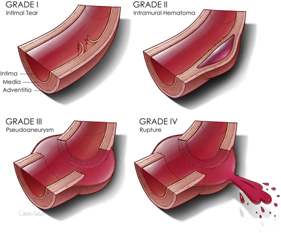
The second paper was presented as a quick-shot at last year’s AAST meeting. It is from a group of researchers from our big Boston trauma centers and the Netherlands. They used four years of data from the TQIP database, giving them extra information unavailable in the first study. They specifically looked at patients with grade II or III injuries. Here is the grading scale:

Here are the factoids:

  • A total of 1,339 patients were studied, with about three-quarters in the early TEVAR group
  • Median time to TEVAR was 4 hours in the early group and 65 hours in the late group
  • Patients in the early group were significantly less likely to have brain or liver injuries
  • ISS was similar in both groups
  • The early TEVAR group had significantly higher in-hospital mortality (16% vs. 5%), significantly higher risk of ARDS (7.6% vs. 2.1%), but significantly shorter ICU stay (7 vs 10 days)
  • When patients who died within the first 24 hours were excluded, the in-hospital mortality remained significantly higher, and the ICU and hospital lengths of stay were significantly shorter

Bottom line: Some society guidelines began recommending delayed TEVAR in 2015. This study did not detect any trend toward this, however. Using different methods and databases, these two studies identified nearly identical mortality and ICU trends in large groups of patients. The mortality trends do not appear to be related to injury grade, overall injury severity, or the presence of head injury. 

Taken together, this suggests that we need to rethink the timing of TEVAR in patients with grade II or III injuries. The best timing still needs to be defined, but it appears to be beyond 24 hours. Centers performing this procedure should review their results and consider extending procedure timing as additional research is done to define the ideal time interval.

References:

  1. Early Versus Delayed Thoracic Endovascular Aortic Repair for Blunt Traumatic Aortic Injury: A Systematic Review and Meta-Analysis. Cureus. 2023 Jun 28;15(6):e41078. doi: 10.7759/cureus.41078. PMID: 37519486; PMCID: PMC10375940.
  2. Early Versus Delayed Thoracic Endovascular Aortic Repair for
    Blunt Thoracic Aortic Injury: A Propensity Score-Matched Analysis. Ann Surg 278:e848-e854, 2023.