Best Of EAST #1: When Is MTP Blood Use Too Much?

The 35th Annual Scientific Assembly of the Eastern Association for the Surgery of Trauma (EAST) begins in only a month! I will be there, sitting in the front row listening to all the great presentations. As usual, I have selected some of the abstracts that I find most interesting and will be sharing my thoughts on them with you over the coming weeks.

Let’s start out with a paper about the massive transfusion protocol (MTP). Blood has always been a scarce resource. And now, thanks to COVID, it is becoming even more so. Every trauma professional reading this has likely been involved in a trauma resuscitation that has used dozens of units of blood and other products. Unfortunately, most of the patients who require this much do not survive.

How does one balance the rapid use of many, many units of blood products with the (un)likelihood of survival and the impact of having less blood for other patients in your hospital or future incoming trauma patients? In other words, when does the use of additional blood become futile? Until now, there have been no real answers to these questions.

The trauma group at George Washington University did a deep dive into the TQIP database seeking some guidance on this topic. They reviewed five years of data, targeting patients who received at least one unit of blood within four hours of arrival. Four-hour and 24-hour mortality was analyzed to determine the point at which additional blood products did not improve survival.

The authors looked at the data two ways. They analyzed the results for all comers, as well as for patients who received balanced resuscitation. Balanced was defined as a red cell to plasma ratio in the range of 1:1 to 2:1. Results were controlled as best as possible for age, sex, race, highest AIS in each body region, comorbidities, advanced directives, and the type of surgery performed to control bleeding.

Here are the factoids:

  • Nearly 100,000 patient records were analyzed, and about 30,000 patients were found to have balanced resuscitation
  • In the all-comers group, mortality plateaued after 41 units at 4 hours and 53 units at 24 hours
  • In the balanced resuscitation patients, mortality plateaued at 40 units (4 hours) and 41 units (24 hours)

The authors concluded that this data should be used as markers for resuscitative timeouts to assess the plan of care.

My comments: This paper is very focused and provides some apparently straightforward results. However, it required some sophisticated statistical analysis to sift through the many variables that need to be controlled to obtain meaningful results. From reading the abstract, it appears that they did a good job of this.

I believe the lower number of units needed by 24 hours in the balanced resuscitation group demonstrates the benefit of getting the MTP ratios right. Non-balanced resuscitation is less efficient / effective and requires the use of more products to hit the mortality plateau.

This paper supports my opinion that a resuscitation timeout is a useful tool in helping us protect our valuable blood product resources and ensuring availability for as many patients in need as possible. What would this look like? Here are my thoughts:

  • Assign one person to monitor the MTP process in real-time. This obviously cannot be the surgeon or a member of the anesthesia team. Or even the operating room crew, as everyone will be very busy. The best practice I’ve seen is to have a dedicated trauma nurse or APP in the ED/OR recording the process on a specialized form and directing which units to give to keep the resuscitation balanced.
  • Call a timeout when the magic threshold is reached. This paper suggests that 40 is a good number.
  • Require that another trauma surgeon come into the room and review the patient condition, operative findings, and progress thus far. The two surgeons should then come to a consensus regarding utility vs futility of further surgery. Based on that decision, the operative procedure either continues or stops.
  • If the operation is to continue, then more timeouts should occur after a defined number of additional products are given.

Here are my questions for the authors / presenter:

  • The statistical analysis required is fairly advanced. Please explain in simple language why the specific regression analysis with bootstrapping was selected.
  • How do you envision applying the thresholds discovered in your paper?

This is an exciting paper and provides important information about the MTP process. I’m looking forward to hearing it in person!

Reference: CRESTING MORTALITY: DEFINING A PLATEAU IN ONGOING MASSIVE TRANSFUSION, EAST 25th ASA, oral abstract #14.

Retained Hemothorax: The Practice Guideline

Over the last few days, I’ve reviewed some data on managing hemothorax, as well as the use of lytics. Then I looked at a paper describing one institution’s experience dealing with retained hemothorax, including the use of VATS. But there really isn’t much out there on how to roll all this together.

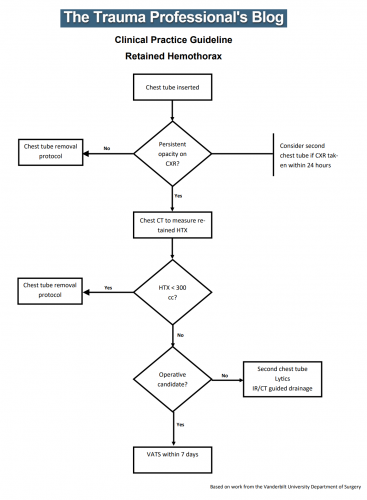
Until now. The trauma group at Vanderbilt published a paper describing their experience with a home-grown practice guideline for managing retained hemothorax.  Here’s what it looks like:

I know it’s small, so just click it to download a pdf copy. I’ve simplified the flow a little as well.

All stable patients with hemothorax admitted to the trauma service were included over a 2.5 year period. The practice guideline was implemented midway through this study period. Before implementation, patients were treated at the discretion of the surgeon. Afterwards, the practice guideline was followed.

Here are the factoids:

  • There were an equal number of patients pre- and post-guideline implementation (326 vs 316)
  • An equal proportion of each group required an initial intervention, generally a chest tube (69% vs 65%)
  • The number of patients requiring an additional intervention (chest tube, VATS, lytics, etc) decreased significantly from 15% to 9%
  • Empyema rate was unchanged at 2.5%
  • Use of VATS decreased significantly from 8% to 3%
  • Use of catheter guided drainage increased significantly from 0.6% to 3%
  • Hospital length of stay was the same, ranging from 4 to 11 days (much shorter than the lytics studies!)

Bottom line: This is how design of practice guidelines is supposed to work. Identify a problem, typically a clinical issue with a large amount of provider care variability. Look at the literature. In general, find it of little help. Design a practical guideline that covers the major issues. Implement, monitor, and analyze. Tweak as necessary based on lessons learned. If you wait for the definitive study to guide you, you’ll be waiting for a long time.

This study did not significantly change outcomes like hospital stay or complications. But it did decrease the number of more invasive procedures and decreased variability of care, with the attendant benefits from both of these. It also dictates more selective (and intelligent) use of additional tubes, catheters, and lytics. 

I like this so much that I’ve incorporated parts of it into the chest tube guideline at my center!

Download the practice guideline here.

Related posts:

Reference: Use of an evidence-based algorithm for patients with traumatic hemothorax reduces need for additional interventions. J Trauma 82(4):728-732, 2017.

Surgical Management Of Retained Hemothorax – VATS

I’ve written about the use of lytics to treat retained hemothorax over the past few days. Although it sounds like a good idea, we just don’t know that it works very well. And they certainly don’t work fast. Lengths of stay were on the order of two weeks in both studies reviewed.

The alternative is video assisted thoracoscopic surgery (VATS). So let’s take a look at what we know about it. This procedure is basically laparoscopy of the chest. A camera is inserted, and other ports are added to allow insertion of instruments to suck, peel, and scrape out the hemothorax.

A prospective, multi-center study was performed over a 2 year period starting in 2009. Twenty centers participated, contributing data on 328 patients with retained hemothorax. This was defined as CT confirmation of retained blood and clot after chest tube placement, with evidence of pleural thickening.

Here are the factoids:

  • 41% of patients had antibiotics given for chest tube placement (this is interesting given the lack of consensus regarding their effectiveness!)
  • A third of patients were initially managed with observation, and most of them (82%) did not need any further procedures (83 of 101 patients)
  • Observation was more successful in patients who were older, had smaller hemothoraces (<300cc), smaller chest tubes (!!, <34 Fr), blunt trauma, and peri-procedure antibiotics (?)
  • An additional chest tube was inserted in 19% of patients, image guided drain placement in 5%, and lytics in 5%. Half to two-thirds of these patients required additional management.
  • VATS was used in 34% of patients. One third of them required additional management including another chest tube, another VATS, or even thoracotomy.
  • Thoracotomy was most likely required if there was a diaphragm injury or large hemothorax (<900cc)
  • Empyema and pneumonia were common (27% and 20%, respectively)

Bottom line: There’s a lot of data in this paper. Most notably, many patients resolve their hemothorax without any additional management. But if they don’t, additional tubes, guided drain placement, and lytics work only a third of the time and contribute to additional time in the hospital. Even VATS and thoracotomy require additional maneuvers 20-30% of the time. And infectious complications are common. This is a tough problem!

Tomorrow, I’ll try to roll it all together and suggest an algorithm to try to optimize both outcomes and cost.

Reference: Management of post-traumatic retained hemothorax: A prospective, observational, multicenter AAST study. 72(1):11-24, 2012.

Retained Hemothorax Part 2: Lytics (again)

In my last post, I reviewed a small case report that was published a couple of years ago on lytics for treatment of retained hemothorax. But surely, there must be something better, right?

After digging around, I did find a paper from 2007 that prospectively looked at protocolized management of retained hemothorax, and its aftermath. It was carried out at a busy Level I trauma center over a 16 month period.

All patients with a hemothorax treated with chest tube received daily chest x-rays. Those with significant opacification on day 3 underwent CT scan of the chest. If more than 300 cc of retained blood was present, the patient received streptokinase or urokinase (surgeon preference and drug availability) daily, and rolled around in bed for 4 hours to attempt to distribute it. The chest tube was then unclamped and allowed to drain. This was repeated for 3 days, and if there was still opacification, a repeat CT was obtained. If the volume was still greater than 300 cc, the cycle was repeated for the next 3 days. If the opacification cleared at any point, or the repeat CT showed less than 300 cc, the protocol was stopped and the chest tube removed. If the chest was still opacified after 6 days, VATS was offered.

Here are the factoids:

  • A total of 203 patients with hemothorax were admitted during the study period and 25 (12%) developed a retained hemothorax
  • While a few had treatment start within 4 days, the majority did not receive lytics until day 9 (range 3  –30 days!)
  • The average length of time in hospital after start of lytics was 7 days, leading to a total length of stay of 18 days
  • 92% of patients had “effective” evacuation of their retained hemothorax, although 1 had VATS anyway which found only 100 cc of fluid
  • 16 patients had “complete” evacuation, and 5 had “partial” evacuation
  • There were no hemorrhagic complications, but one third of patients reported significant pain with drug administration

Bottom line: Sounds good, right? The drug seems reasonably effective, although lengths of stay are relatively long. However, streptokinase and urokinase are no longer available in the US, having been replaced with tissue plasminogen activator (tPA). This paper does a cost analysis of lytics vs VATS and found that the former treatment cost about $15000 (drug + hospital stay) vs $34000 for VATS. However, a big part of this was that the drug only cost about $75 per dose. tPA is much more expensive.

So once again, small series, longer lengths of stay, but at least nicely done. Unfortunately, the drug choice is no longer available so use of tPA tilts the balance away from lytics. 

Reference: Intrapleural Thrombolysis for the Management of Undrained Traumatic Hemothorax: A Prospective Observational Study. J Trauma 62(5):1175-1179, 2007.

Retained Hemothorax Part 1: Lytics

In my last pair of posts, we reviewed chest tube management. Now let’s dive into hemothorax. This gets a little complicated, because a chest tube doesn’t always do what it’s supposed to when it comes to blood and possible blood clots.

Hemothorax is a common complication of chest trauma, occurring in about one third of cases. It is commonly treated with a chest tube, which usually takes care of the problem. But in a few cases some blood remains, which can result in an entrapped lung or empyema.

There are several management options. Historically, these patients underwent thoracotomy to peel out the fibrinous collection stuck to lung and chest wall. This has given way to the more humane VATS procedure (video assisted thoracoscopic surgery) which accomplishes the same thing using a scope. In some cases, another tube can be inserted, sometimes under CT guidance, to try to drain the blood.

So what about lytics? It’s fibrin, right? So why not just dissolve it with tissue plasminogen activator (tPA)? There have been very few studies published over the years. The most recent was in 2014. I’ll review it today, and another tomorrow. Finally, I’ll give you my thoughts on the best way to deal with retained hemothorax.

Here are the factoids:

  • This was a single center, retrospective review of data from 1.5 years beginning in 2009
  • A total of seven patients were identified, and most had hemothorax due to rib fractures. Three presented immediately after their injury, 4 were delayed.
  • Median time from injury to chest tube placement was 11 days
  • Median time the chest tube was in place was 13 days, with an average hospital stay of 14 days
  • Patients received 1 to 5 treatments, averaging 24mg per dose
  • There was one death in the group, unrelated to TPA treatment
  • No patient “required” VATS, but one underwent thoracotomy, which turned out to be for a malignancy

Bottom line: The authors conclude that tPA use for busting retained hemothorax is both safe and effective. Really? With only seven patients? The biggest problem with this study is that it uses old, retrospective data. We have no idea why these patients were selected for tPA in this 5-year old cohort of patients. Why did it take so long to put in chest tubes? Why did the chest tubes stay in so long? Maybe this is why they were in the hospital so long?

Plus, tPA is expensive. A 100mg vial runs about $6000. Does repeatedly using an expensive drug and keeping a patient in the hospital an extra week or so make financial sense? So it better work damn well, and this small series doesn’t demonstrate that.

Tomorrow, I’ll look at the next most recent paper on the topic, from way back in 2004.

Reference: Evaluation of chest tube administration of tissue plasminogen activator to treat retained hemothorax. Am J Surg 267(6):960-963, 2014.