Does Time To Interventional Radiography Make a Difference In Solid Organ Injury?

Solid organ injury is one of the more common manifestations of blunt abdominal trauma. Most trauma centers have some sort of practice guideline for managing these injuries. Frequently, interventional radiology (IR) and angioembolization (AE) are part of this algorithm, especially when active bleeding is noted on CT scan.

So it makes sense that getting to IR in a timely manner would serve to stop the bleeding sooner and help the patient. But in most hospitals, interventional radiology is not in-house 24/7. Calls after hours require mobilization of a call team, which may be costly and take time.

For this reason, it is important to know if rapid access to angioembolization makes sense. Couldn’t the patient just wait until the start of business the next morning when the IR team normally arrives?

The group at the University of Arizona at Tucson tackled this problem. They performed a 4-year retrospective review of the TQIP database. They included all adult patients who underwent AE within four hours of admission. Outcome measures were 24-hour mortality, blood product usage, and in-hospital mortality.

Here are the factoids:

  • Out of over a million records in the database, only 924 met the inclusion criteria
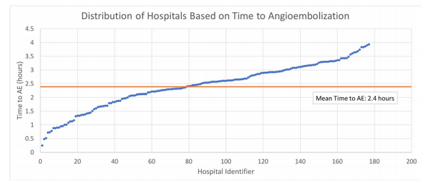
  • Mean time to AE was 2 hours and 22 minutes, with 92% of patients getting this procedure more than an hour after arrival
  • Average 24-hour mortality was 5%. Mortality by hours to AE was as follows:
    • Within 1 hour: 2.6%
    • Within 2 hours: 3.6%
    • Within 3 hours: 4.0%
    • Within 4 hours: 8.8%
  • There was no difference in the use of blood products

The authors concluded that delayed angioembolization for solid organ injury is associated with increased mortality but no increase in blood product usage. They recommend that improving time to AE is a worthy performance improvement project.

Bottom line: This study has the usual limitations of a retrospective database review. But it is really the only way to obtain the range of data needed for the analysis. 

The results seem straightforward: early angioembolization saves lives. What puzzles me is that these patients should be bleeding from their solid organ injury. Yet longer delays did not result in the use of more blood products.

There are two possibilities for this: there are other important factors that were not accounted for, or the sample size was too small to identify a difference. As we know, there are huge variations in how clinicians choose to administer blood products. This could easily account for the apparent similarities between products given at various time intervals to AE.

My advice? Act like your patient is bleeding to death. If the CT scan indicates that they have active extravasation, they actually are. If a parenchymal pseudoaneurysm is present, they are about to. So call in your IR team immediately! Minutes count!

Reference: Angioembolization in intra-abdominal solid organ injury:
Does delay in angioembolization affect outcomes?  J Trauma 89(4):723-729, 2020.

In The Next Trauma MedEd Newsletter: Fatigue And Sleep Loss

The next issue of Trauma MedEd will be sent out to subscribers this week, and will provide some interesting information on fatigue and sleep loss.

Most trauma professionals have at least a little experience with this topic. You may one who provides care on call at night, or work a crazy shift system. We all end up on the short end of the sleep stick at times.

Topics will include:

  • Facts on fatigue and sleep loss
  • Impact on EMS providers
  • Impact of Nurses
  • Impact on Physicians and APPs
  • What to do about it

As always, this month’s issue will go to all of my subscribers first. If you are not yet one of them, click this link right away to sign up now and/or download back issues.

Unfortunately, non-subscribers will have to wait until I release the issue on this blog, in mid-June. So sign up now!

How To Detect Bucket-handle Intestinal Injuries With CT Scan

A bucket-handle injury is a relatively uncommon complication of blunt trauma to the abdomen. It only occurs in a few percent of patients, but is much more likely if they have a seat belt sign.  The basic pathology is that the bowel mesentery (small bowel of sigmoid colon) gets pulled away from the intestinal wall.

This injury is problematic because it may take a few days for the bowel itself to die and perforate. Patients with no other injuries could potentially be discharged from the hospital before they become overtly symptomatic, leading to delayed treatment.

Here’s an image from my personal collection with not one, but four bucket-handle injuries.

Typical patients with suspected blunt intestinal injury are observed with good serial exams and a daily WBC count. If this begins to rise after 24 hours, there is a reasonable chance that this injury is present.

CT scan has not really been that reliable in past studies. There may be some “dirty mesentery”, which is contused and has a hematoma within it. But without a more convincing exam, it is difficult to convince yourself to operate immediately on these patients.

A paper was published by a group of radiologists at Duke University. It appears to be a case report disguised as a descriptive paper. It looks like they picked a few known bucket-handle injuries from their institution and back-correlated them with CT findings.

The authors called out the usual culprits:

  • Fluid between loops of bowel
  • Active bleeding in the mesentery
  • Bowel wall perfusion defects

But they also noted that traumatic abdominal wall hernias were highly with the injury as well. These are rare, but should bring intestinal injury to mind when seen.

With newer scanners, radiologists are better able to detect subtle areas of hypoperfusion as well. This is a fairly good indicator of injury, especially when adjacent bowel appears normally perfused. Here are two examples. The black arrows denote active extravasation, and the white ones an area of hypoperfusion.

The authors add bowel wall hypoperfusion as another finding that may point to a bucket-handle type injury

Bottom line: Hold the phone! Don’t change your practice yet. This paper is not able to demonstrate how good this radiographic sign is. Looking at other radiology literature, the specificity is about 90%. But remember, that means that if they don’t have the CT finding, that’s true only 90% of the time.

Unfortunately the sensitivity is only 10%. So if you see it on the scan, they’ve got a 1 in 10 chance of actually having the injury. That’s not good enough for me to run to the operating room.

Here’s what I recommend: if your patient has an unconcerning exam and any of the usual culprits (pelvic fluid, inter-loop fluid, dirty mesentery, thickened bowel loops, abdominal wall hernia), perform serial exams and get a WBC the next morning. If the exam worsens, operate. If the WBC rises, consider laparoscopy to see if you need to make a bigger incision. And if you see this new kid on the block, the hypoperfused bowel, consider laparoscopy right away. 

I’m sure the radiologists and the technology will keep getting better. But for now, blunt intestinal injury still requires patience, perceptiveness, and a little luck.

References:

  • CT findings of traumatic bucket-handle mesenteric injuries. Am J Radiol 209:W360-@364, 2017.
  • Multidetector CT of blunt abdominal trauma. Radiology 265(3):678–693, 2012.

What Is A Wide Mediastinum Anyway?

Trauma professionals are always on the lookout for injuries that can kill you. Thoracic aortic injury from blunt trauma is one of those injuries. Thankfully, it is uncommon, but it can certainly be deadly.

One of the screening tests used to detect aortic injury is the old-fashioned chest xray. This test is said to be about 50% sensitive, with a negative predictive value of about 80%. However, the sensitivity is probably decreasing and the negative predictive value increasing due to the rapidly increasing number of obese patients that we see.

A wide mediastinum is defined as being > 8cm in width. In this day and age of digital imaging, you will need to use the measurement tool on your workstation to figure this out.

Unfortunately, it seems like most chest xrays show wide mediastinum these days. What are the most common causes for this?

  • Technique. The standard xray technique used to reduce magnification of the anterior mediastinum (where the aortic arch lives) is a tube distance of 72 inches from the patient, shot back to front. We can’t do this for trauma patients because we can’t stand them up and are reluctant to prone them. The standard trauma room technique is 36 inches from the patient shot front to back. This serves to magnify the mediastinal image and make it look wide.
  • Obesity. The more fat in the mediastinum, the wider it looks. The more fat on the back, the further the mediastinum is from the xray plate and the greater the magnification.
  • Other mediastinal blood. Major blunt trauma to the chest can cause bleeding from small veins in the mediastinum, making it look wide.
  • Thymus. Only in kids, though.
  • Aortic injury. Last but not least. Only a few percent of people with wide mediastinum will actually have the injury.

If you encounter a wide mediastinum on chest xray in a patient with a significant mechanism for aortic injury, then they should be screened using helical CT.

Is The Trauma Bay Chest X-Ray Really Necessary Or Just Dogma?

I love challenging dogma. I spoke last week (virtually) at an excellent event at the Intermountain Medical Center in Utah. One of my talks there addressed trauma myths and dogma.

I bring this up because there is an interesting article in the Journal of Trauma this month that questions the necessity of the routine chest x-ray (CXR) in blunt trauma resuscitation. So of course, this caught my eye. Let’s dig in.

The first thing to understand is that this article is an opinion piece and is identified as such. It was written by three surgeons, including the trauma medical director, at the Stanford University Hospital trauma center.

First, what are we really looking for on the chest x-ray that is taken in the trauma bay? I call them “the three big things”.

  • Big air. The first item to be identified is a pneumothorax. The chest x-ray helps the trauma professionals decide if the pneumo needs an intervention (chest tube) and when. (Note: it could in theory identify a tension pneumothorax. But in that case, the trauma pros should be embarrassed. They should have picked that up on their clinical exam and assessment of the vitals.)
  • Big blood. The chest x-ray can also identify a hemothorax. And once again, it can help decide whether its size warrants chest tube insertion.
  • Big mediastinum. A wide mediastinum may indicate the presence of hematoma from an aortic injury. It is one of the indications for performing CT angiography of the chest to rule it out.

Here are their authors’ arguments:

  • There are other imaging modalities available to us that are very accurate. FAST ultrasound has been used routinely for abdominal and cardiac evaluation for over a decade. The extended FAST (eFAST) involves evaluation of the pleural interface to identify pneumothorax. A study published last year pitted CXR vs eFAST. It found that the eFAST outperformed with a sensitivity of 94% and specificity of nearly 100%.
    But what about hemothorax? Ultrasound is less helpful here. But the CT scanner is. It is far more accurate at identifying and quantifying hemothorax than the CXR.
  • Evaluation of the aorta can either wait, or it can’t wait at all. If the patient loses vital signs in the trauma bay the decision to open the chest or insert a REBOA catheter must be made. In the latter case, a chest x-ray must be obtained to exclude a thoracic source of bleeding that the cathether is of no use for. But if the patient truly is bleeding out from a blunt aortic injury, it is nearly certain that he or she is not leaving the trauma bay alive.
    What about using the wide mediastinum as an indication or order the chest CT angiogram? The authors argue that there will probably be a history of deceleration or other associated injuries (femur fracture is a very common one).

Bottom line: The authors argue that the chest x-ray should go the way of the lateral cervical spine x-ray used at the turn of the 21st century and before. They claim that judicious use of the extended FAST and CT angiography can identify the significant injuries we need to know about in a timely manner.

My own opinion is more nuanced. I buy their arguments that the extended FAST will identify all significant pneumothoraces. However, we have typically answered the question “how big is too big” using the chest x-ray. That is the most helpful tool in deciding whether a chest tube is warranted or not.

As for hemothorax, I don’t believe that a CT is the best tool for evaluating this, either. Are the authors members of the “pan-scan” school? What about those of us that use the “selective scan” philosophy. True, the abdominal scan will identify both hemothorax and pneumthorax on the lower cuts of the chest. But as in the previous paragraph, we are better trained to judge when a chest tube is indicated by the appearance of the chest x-ray. Hemothorax (or pneumothorax) is not an indication to get a chest CT.

I don’t buy argument that there will be other indications of potential aortic injury. Deceleration is in the eye of the beholder. How do we know how fast the vehicle was actually moving? What is the magic velocity that will break this patient’s aorta? This particular patient may not have any of the other potential indicators that increase suspicion for aortic injury. That wide mediastinum may be the only clue. Yes, the numbers of affected patients are small, but the consequences of missing one could be deadly.

And what about patients who might not get scanned at all? And those who need a study to confirm tube or line placement? They must absolutely get a chest x-ray before they leave the trauma bay.

At this point, I can’t see a way to dispense with the chest x-ray completely. It should still be used to:

  • Confirm pneumothorax from eFAST to help decide if a chest tube is needed
  • Identify potential pathology (hemothorax, wide mediastinum) in patients who don’t otherwise meet criteria for chest CT
  • Verify endotracheal tube position after intubation

What do you think? Please leave your comments or Tweets about this topic.

References:

  • Extended-FAST plus MDCT in pneumothorax diagnosis of major trauma: time to revisit ATLS imaging approach? J Ultrasound. 2019;22(4):461–469.
  • Necessity of routine chest radiograph in blunt trauma resuscitation: Time to evaluate dogma with evidence. J Trauma 2020;89(3):e69-70.