Epidural Hematoma Treated With Middle Meningeal Artery Embolization

Epidural hematoma is a life-threatening condition that is typically associated with arterial bleeding outside of the dura. Most frequently, this is due to a skull fracture that extends across and lacerates the middle meningeal artery (MMA).

The standard treatment regimen is neurologic monitoring in patients who have a (nearly) normal GCS and do not change neurologically. That escalates to rapid craniectomy and evacuation in those with neurologic compromise.  Interestingly, there have been a few case reports over the last 10 years describing attempted management by embolization of the MMA.

Let’s look at this idea more critically. This seems like it should be a good idea. But remember, in medicine you’ve actually got to study it. There are too many examples of things that make sense that are worthless or actually cause harm.

The first report I found was a series of one in which the patient was found to have a large subdural hematoma. He was taken to surgery and the lesion was evacuated. However, there was persistent epidural bleeding intraop which was thought to be controlled. Repeat scan the next day showed a large epidural, so he was returned to the OR. Once again, there was persistent epidural oozing and the collection was removed. Followup CT showed yet another epidural. The patient was finally taken to interventional radiology for embolization of the MMA. This was successful, and the patient had no further recurrences.

This case provided proof of concept, although the bleeding was not due to known traumatic injury to the MMA. Last year, another case report was published that described an experience (of one again) with a young male who was found down. He awakened and then became obtunded again. CT showed bilateral epidural hematomas. He was taken to the OR for operative evacuation of the larger one. Postop CT showed expansion of the smaller one.

The patient was then taken to the endovascular suite and MMA embolization was carried out. The hematoma stabilized and the patient was later discharged without sequelae.

This case was trauma-related, but not for an acute bleed. Now, let’s look at a bigger case series to see how well this works. This one detailed the experience of a neurosurgery group in Sao Paulo, Brazil. All patients who underwent conservative management based on “standard criteria” were studied. Patients with large hematomas, midline shift, depressed skull fracture, coagulopathy, or incomplete data were excluded. One third of the injuries were due to falls, and the rest were due to other blunt mechanisms.

Here are the factoids:

  • 85% had an attendant skull fracture
  • About 82% had active extravasation from the MMA
  • All patients had followup CT scan 1-7 days after the procedure, and no increase in epidural size was noted
  • None of the patients had a change in GCS or needed operative intervention
  • The authors compared these results to historical controls from other published literature

Bottom line: Sounds impressive, right? But not so fast, there are a lot of loose ends here. First, these are supposedly all patients with epidural hematoma who were treated without operation. Decision to operate was based on criteria set out in a paper published 15 years ago. This strains the imagination a bit. There is usually no uniformity in the way individual neurosurgeons decide to operate, so it is likely there may be some significant selection bias here. It is very easy to believe that patients who were predicted to do well were the only ones enrolled in the study. This also explains why the authors had to use controls from other authors’ research for outcome comparison.

The results are too clean as well. No adverse events. No patients who ended up needing surgery. Followup scans were performed any time between postop day 1 and 7, but there is no frequency breakdown. If most of the repeat scans were performed near the beginning of the postop period, little change would be expected. MMA embolization is either a miracle cure or …

You know what they say, “if it seems to good to be true…” A single case series like this should never change one’s practice. Middle meningeal artery embolization sounds like common sense, but the devil is always in the details. This concept needs a lot more study before you should ever consider it in your patients. Or, you could start a real, IRB-approved study and make an excellent contribution to the neurosurgery literature.

References:

  1. Embolization of the Middle Meningeal Artery for the Treatment of Epidural Hematoma. J Neurosurg 110(6):1247-1249, 2009.
  2. Middle Meningeal Artery Embolization for the Treatment of an Expanding Epidural Hematoma. World Neurosurg 128:284-286,2019
  3. Endovascular Management of Acute Epidural Hematomas: Clinical Experience With 80 Cases. J Neurosurg 128(4):1044-1050, 2018.

What Is: A Rubber Bullet?

The protests in cities across the country continue. Many are peaceful, but not all. In some cases, police have resorted to “non-lethal” weapons to control and disperse crowds.

Although these weapons are called non-lethal, that’s not entirely true. The projectiles, gases, and powders that are being used all have some degree of morbidity and mortality. They are certainly less so than traditional projectiles (bullets), but serious and fatal injures can and do occur.

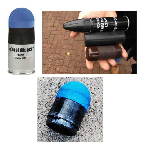
One item that is talked about in the news is the rubber bullet. What are these, exactly? The generic term is a “kinetic impact projectile” (KIP). It encompasses a variety of objects that are not designed to penetrate flesh like a regular bullet. They can be bullets, beanbags, sponges, pellets, and other odds and ends.

And the so-called “rubber bullet” isn’t even necessarily made of rubber. It can be plastic, metal, rubber, or other substances.

There is very little published data on injuries caused by KIPs. Because of their odd shapes, they tend to tumble when they are fired. This decreases aiming accuracy substantially when the target is distant. They are designed to be aimed at the lower extremities. However, if the aim is too high or the round is fired at close range, it can be lethal.

Here are some typical injuries that have been descirbed:

  • Subdural and intraparenchymal hematomas
  • Skull and facial fractures
  • Eye injuries leading to blindness (this happened to a photographer in Minneapolis last week)
  • Rib fractures and pulmonary contusions
  • Spleen laceration
  • Blunt intestinal injury

Here’s a video from one of the manufacturers that shows the amount of target deformation caused by a sponge tipped bullet. Very impressive.

YouTube player

(Tumblr viewers please click here to view video)

Bottom line: Although they sound relatively innocuous, kinetic impact projectiles of any kind are far from it. If you are called to treat a patient who has been shot with one, be sure to do a very thorough evaluation. Most head, neck, and torso injuries should undergo CT scanning to delineate deep or occult injuries in detail. In-hospital observation of torso injuries is warranted, as well as a good tertiary exam.

More On Lead Poisoning And Retained Bullets

Trauma professionals frequently have to leave bullets in patients. It is often more disruptive to go digging the projectiles out than to just leave them in place. But patients always want to know why and what the consequences might be.

In my last post, I discussed a very old paper on what we know about lead levels and retained bullets. Very recently, a meta-analysis was published that provides a better picture of this topic. They somehow managed to find over 2000 articles dealing with lead toxicity and bullets out there. But after someone had the pleasure of reviewing each of them, they found only 12 that had any meaningful or actionable information.

Here are the factoids:

  • All studies were observational (duh! It would be difficult to get your IRB to approve a study where patients were shot on purpose)
  • There were five cross-sectional studies, four case-control studies, and three prospective cohort studies
  • The studies were small, with a median of only 26 patients (range 15-120)
  • Eleven of the twelve studies showed an association with retained bullets and elevated blood lead levels
  • Three studies showed elevated blood levels if a fracture was present
  • The higher the number of retained fragments, the more likely lead levels were to be high
  • Higher lead levels were associated with retained fragments near a bone or joint
  • There were no good correlations with number of fragments and location vs actual lead toxicity

Bottom line: Even using meta-analysis, it is difficult to tease out meaningful answers to this question. That speaks to the low numbers of papers and their quality. However, this study does provide a little bit of guidance.

Retained bullet fragments are probably not a big worry in most patients. The bothersome cases are those where the fragments are in or near a bone or joint. And even though few patients actually developed lead toxicity, lead levels approaching 5 micrograms/dL can have physiologically significant negative effects. 

Recommendation: If your patient has a retained bullet fragment near a bone or joint, or they have “multiple” retained fragments (no good definition of this), they should have blood lead levels measured every three months for a year. If the level is rising, and certainly if it reaches the 5μ/dL level, attempts should be made to remove the fragments.

Reference: Lead toxicity from retained bullet fragments: A systematic review and meta-analysis. J Trauma 87(3):707-716, 2019.

Can Lead Poisoning Occur After A Gunshot?

This is a fairly common question from victims of gunshots and their families. As you know, bullets are routinely left in place unless they are superficial. It may cause more damage to try to extract one, especially if it has come to rest in a deep location. But is there danger in leaving the bullet alone?

One of the classic papers on this topic was published in 1982 by Erwin Thal at Parkland Hospital in Dallas. The paper recounted a series of 16 patients who had developed signs and symptoms of lead poisoning (plumbism) after a gunshot or shotgun injury. The common thread in these cases was that the injury involved a joint or bursa near a joint. In some cases the missile passed through the joint/bursa but came to rest nearby, and a synovial pseudocyst formed which included the piece of lead. The joint fluid bathing the projectile caused lead to leach into the circulation.

The patients in the Parkland paper developed symptoms anywhere from 3 days to 40 years after injury. As is the case with plumbism, symptoms were variable and nonspecific. Patients presented with abdominal pain, anemia, cognitive problems, renal dysfunction and seizures to name a few.

Bottom line: Any patient with a bullet or lead shot that is located in or near a joint or bursa should have the missile(s) promptly and surgically removed. Any lead that has come to rest within the GI tract (particularly the stomach) must be removed as well. If a patient presents with odd symptoms and has a history of a retained bullet, obtain a toxicology consult and begin a workup for lead poisoning. If levels are elevated, the missile must be extracted. Chelation therapy should be started preop because manipulation of the site may further increase lead levels. The missile and any stained tissues or pseudocyst must be removed in their entirety.

Granted, this is a very old paper. Over the years, a few papers on the topic have popped up from time to time. In my next post, I’ll review a meta-analysis on this topic that was published just last year.

Reference: Lead poisoning from retained bullets. Ann Surg 195(3):305-313, 1982.

Fractures From Gunshots: Open Fracture Or Not?

Penetrating trauma has been increasing over recent years, especially here in sleepy St. Paul MN. On occasion, we all see patients who have sustained gunshots that have caused fractures. The persistent question has been: open fracture or not?

Do these patients need antibiotics? A wound washout? Are they at risk for lead poisoning? Unfortunately, there are no consistent answers in the textbooks. The orthopedic trauma group and MetroHealth in Cleveland sent surveys to 385 members of the Orthopedic Trauma Association (OTA) to see if there was some consensus.

A total of 173 of the surveys were completed, which is actually a very good success rate.  About 72% were in practice at a Level I center, 18% at a Level II, and 10% at Level III/IV or non-trauma centers.

There was considerable heterogeneity among the responses. Here are the summaries for the specific questions asked:

How would you treat a gunshot injury near bone without fracture?

The majority of respondents recommended non-operative treatment and some form of antibiotics. However, there was no consensus regarding route of administration or duration. About 75% were in favor of a single dose of IV antibiotics, and half of those also recommended addition oral antibiotics. The presence of a retained bullet did not change management.

How would you treat a gunshot with a stable fracture to the fibula?

Three quarters of the respondents recommended the same management as above (IV antibiotics + oral), although about 10% would admit for IV antibiotics and 10% would do a washout or debridement. Only 7% recommended no antibiotics or debridement.

How would you treat a gunshot traversing the  knee joint with a retained bullet?

About half stated they would explore the joint and the other half would not. Nearly all recommended antibiotics, with the majority in favor of a single dose IV followed by some duration of oral.

Is the union rate of a tibial shaft fracture from a gunshot treated surgically different than a non-gunshot fracture?

Half of the participants thought it would be the same, a quarter thought it would be higher, and a quarter lower.

What about a gunshot with a displaced tibia fracture without other skin wounds?

About half recommended fixation with irrigation and debridement with perioperative antibiotics. A quarter would do the same, but without the irrigation and debridement. About 10% would extend the antibiotic duration.

How would you handle a gunshot traversing bowel that results in a stable pelvic fracture?

There was no agreement here at all. The majority (61%) would not debride the fracture, but would recommend IV antibiotics. Most of those recommended at least 24 hours of coverage. The remaining surgeons recommended surgical debridement, and were evenly split over brief vs longer antibiotic duration.

Bottom line: This is a “How we do it study” that is based on science as interpreted by these orthopedic surgeons. In general, OTA members behave as if they consider gunshots to bone as open fractures. More than 90% recommend antibiotics any time a bullet touches the bone. But once the fracture requires operative management, it is treated like a non-gunshot fracture from the standpoint of debridement and antibiotics.

The most interesting part of this survey was the total lack of consistency in the answers. It is clear that there is wide variation in the practice patterns of these surgeons, which usually signifies a lack of good data pertaining to the problem.

In my next post, I’ll discuss the lead poisoning question I mentioned above.

Reference: Variation in treatment of low energy gunshot injuries – a survey of OTA members. Injury 49:570-574, 2018.