Inserting an NG Tube (Not an NC Tube)!

On occasion (but not routinely) trauma patients need to have their stomach decompressed. The reflex maneuver is to insert a nasogastric (NG) tube. However, this may be a dangerous procedure in some patients.

Some patients may be at risk for a cribriform plate fracture, and blindly passing a tube into their nose may result in a nasocerebral (NC) tube (see picture). Immediate and profound neurologic decompensation usually occurs. This is a neurosurgical catastrophe, and the outcome is uniformly dismal. It generally requires craniectomy to remove the tube.

The following patients are at risk:

  • Evidence of midface trauma (eyebrows to zygoma)
  • Evidence of basilar skull fracture (raccoon eyes, Battle’s sign, fluids leaking from ears or nose)
  • Coma (GCS<8)

If you really need the tube, what can you do? If the patient is comatose, it’s easy: just insert an orogastric (OG) tube. However, that is not an option in awake patients; they will continuously gag on the tube. In that case, lubricate a curved nasal trumpet and gently insert it into the nose. The curve will safely move it past the cribriform plate area. Then lubricate a smaller gastric tube and pass it through the trumpet.

Flash Pulmonary Edema After Chest Tube Insertion

You are seeing a young man in the emergency department who gives a history of falling two days ago. He experienced chest pain at the time which has persisted, but he did not immediately seek medical care. He has noticed that he now gets winded when walking quickly or climbing stairs, and describes pleuritic chest pain.

He presents to your emergency room and on exam has a bruise over his left lateral chest wall. Subcutaneous emphysema is present, and breath sounds are absent. Chest x-ray shows a complete pneumothorax on the left.

You carefully prepare and insert a chest tube in the usual position. A significant rush of air occurs, which tapers off over 15 seconds. Here is the followup image:

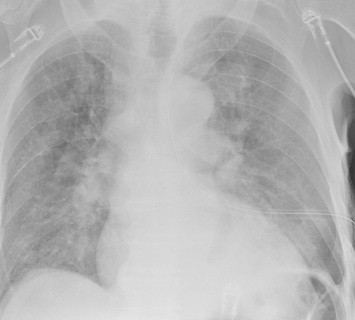
About 10 minutes later you are called to his room because he is complaining of dyspnea and his oxygen saturation has decreased to 86%. Breath sounds are somewhat decreased and the tube appears to be functioning properly. You immediately obtain another chest x-ray:

What just happened? This is a classic case of unilateral “flash” pulmonary edema after draining the chest cavity. This phenomenon was first described in 1853 in a patient who had just undergone thoracentesis. It is very uncommon, but seems to occur after rapid drainage of air or fluid from the chest cavity.

Here are some interesting factoids from case reports:

  • It occurs more often in young men
  • It is most common when draining large hemo- or pneumothoraces
  • Rapid drainage seems to increase the incidence
  • It is likely due to increased pulmonary capillary permeability from inflammatory mediators or changes in surfactant
  • Symptoms typically develop within an hour after drainage

What should you do? First, if you are draining a large collection of air or blood, do it slowly. Clamp the back end of the chest tube prior to insertion (you should always do this if you value your shoes) and use it to meter the amount of fluid or air released. I typically let out about 300cc of fluid, then wait a minute and repeat until all the blood has been drained. For air, vent it for 10 seconds, then wait a minute and repeat.

In patients at high risk for this condition, apply pulse oximetry and follow for about an hour. If they still look and feel great, nothing more need be done.

References:

  • Fulminant Unilateral Pulmonary Edema After Insertion of a Chest Tube. Dtsch Arztebl Int 105(50):878-881, 2008.
  • Reexpansion pulmonary edema after chest drainage for pneumothorax: A case report and literature overview. Respir Med Case Rep 14:10-12, 2015.
  • Re-expansion pulmonary edema following thoracentesis, Can Med Assn J 182(18):2000-2002, 2010.

What Is: A Pars Fracture / Defect

Radiologists sure know their anatomy! The vast majority of the time, I actually know what they are describing. But every once in a while they’ll toss in some term that I know I probably learned about in medical school (last century). For whatever reason though, I’m just not able to retrieve it.

Which brings me to the pars fracture. Hmm. I figure that if I have to hit the books again to look something up, there are probably a few other trauma professionals out there who are dying to know what it is, too. Here’s a diagram of a typical vertebra:

The arch extending away from the vertebral body consists of the pedicles, which are connected by the lamina. A number of things jut off from this arch, including the transverse and spinous processes and the articular processes.

The area between the lamina and pedicle and adjacent to the articular process is called the pars interarticularis. This area is a bit thinner and flatter than the rest of the arch and can fracture if sufficient acute stress is applied. It can also fracture if enough chronic stress in the area occurs. This pattern is typically seen in the lumbar spine, but may also occur at the cervical level. Thus, a pars fracture or pars defect is simply a fracture through this area.

Another term you may see with regard to the pars is spondylolysis. This is defined as a defect in the pars interarticularis, typically from a fracture. So if you see either of these terms in a radiology report, recognize that they are basically one and the same.

Here is a nice image showing the location of the pars, and the axial CT appearance of “bilateral pars defects.”

Mystery solved! Amaze your friends!

The Cost Of Duplicate Radiographic Studies

Speaking of radiation, here’s another tidbit. Duplicate radiographic studies are a continuing issue for trauma professionals, particularly after transfer from a smaller hospital to a trauma center. The incidence has been estimated anywhere from 25% to 60% of patients. A lot has been written about the radiation dangers, but what about cost?

A Level II trauma center reviewed their experience with duplicate studies in orthopedic transfer patients retrospectively over a one year period. They looked at the usual demographics, but also included payor, cost information, and reason for repeat imaging. Radiation dose information was also collected.

Here are the factoids:

  • 513 patients were accepted from 36 referring hospitals
  • 48% had at least one study repeated, 256 CT scans and 161 conventional imaging studies
  • Older patients and patients with low GCS were much more likely to receive repeat studies
  • There were no association with the size of the referring hospital or the ability of the patient to pay
  • Most transfers had commercial insurance; only 11% had Medicaid and 17% were uninsured
  • Additional radiation from repeat scans was 8 mSv. The average radiation dose from both hospitals was 38 mSv. This is 13 years of background radiation exposure!
  • The cost of all the repeat studies was over $96,000

Bottom line: This is an eye-opening study, particularly regarding how often repeat imaging is needed, how much additional radiation is delivered, and now, the cost. And remember that these are orthopedic patients, many of whom had isolated bony injuries. I would expect that patients with multiple and multi-system injuries would require more repeat imaging and waste even more money. It is imperative that all centers that receive transfers look at adopting some kind of electronic data transfer for imaging, be it a VPN or some cloud-based service. With the implementation of the Orange Book by the American College of Surgeons, Level I and II centers will receive a deficiency if they do not have some reliable mechanism for this.

“Level I and II facilities must have a mechanism in place to view radiographic imaging from referring hospitals within their catchment area (CD 11–42).”

Reference: Clinical and Economic Impact of Duplicated Radiographic Studies in Trauma Patients Transferred to a Regional Trauma Center. J Ortho Trauma 29(7):e214-e218, 2015.

How Much Radiation Exposure In Imaging Studies?

Everyone knows that CT scans deliver more radiation than conventional x-ray. But how much does each test really deliver? And how significant is that?

Let me try to put it all into perspective. First, how much radiation are we exposed to just living outside the hospital? Background radiation is everywhere. It consists of radioactive gases (argon) in the air we breathe, radiation from the rocks and other things around us, and cosmic rays blasting through us from space.

In the United States, the average background radiation each of us is exposed to is about 3.1 milliSieverts (mSv). I’ve compiled a table to show the approximate dose delivered by some of the common radiographic studies ordered by trauma professionals. And to keep it real, I’ve calculated how much extra background radiation we would have to absorb, in units of time, to have an equivalent exposure.

Read and enjoy! Remember, doses may vary by scanner, settings, and dose reduction measures used.

Test Dose (mSv) Equivalent background
radiation
Chest x-ray 0.1 10 days
Pelvis x-ray 0.1 10 days
CT head 2 8 months
CT cervical spine 3 1 year
Plain c-spine 0.2 3 weeks
CT chest 7 2 years
CT abdomen/pelvis 10 3 years
CT T&L spine 7 2 years
Plain T&L spine 3 1 year
Millimeter wave
scanner (that hands
in the air TSA thing at
the airport)
0.0001 15 minutes
Scatter from a chest
x-ray in trauma bay
when standing one 
meter from the
patient
0.0002 45 minutes
Scatter from a chest
x-ray in trauma bay
when standing three 
meters from the
patient
0.000022 6 minutes