What Is: The Monteggia Fracture

Yesterday, I wrote about one of the many fractures that occurs during falls onto outstretched hands, the Galeazzi fracture. Today, I’ll describe another one, the Monteggia fracture. Yes, this one is named after another Italian surgeon! And like the other one, the person it was named after was actually the second to describe it.

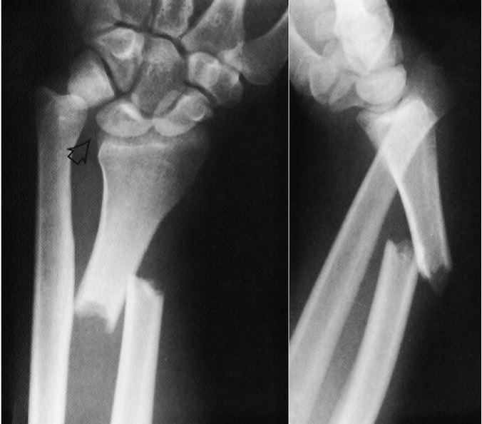
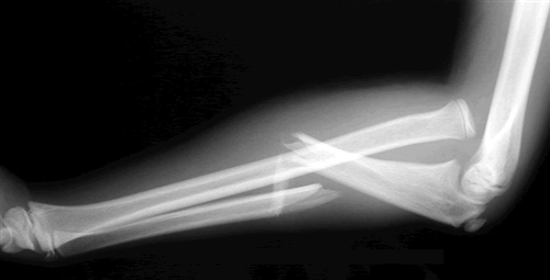
Think of the Monteggia fracture as the exact opposite of a Galeazzi fracture. The fractured bone is switched, as is the dislocation. Whereas the Galeazzi is a distal radius fracture with a distal ulnar dislocation which pulls the radio-ulnar joint apart, the Monteggia is a proximal ulnar fracture with a proximal radial head dislocation.

Here’s what it looks like:

Of course, the orthopedic surgeons have a classification system for this based on the directions the bones fracture and dislocate. I won’t bore you with the details.

Unlike the Galeazzi fracture, all of these require operative repair, even in children. This helps stabilize the radial head and decreases the incidence of malunion.

What Is: The Galeazzi Fracture?

Orthopedic surgeons have so many names for fractures, it gets confusing! Today, let’s dig in to the “Galeazzi fracture.” This one was named for an Italian surgeon during the early 20th century) although it was actually first described by an Englishman named Cooper a hundred years earlier).

The Galeazzi fracture is an uncommon one, and consists of two components: a radius fracture at the junction of the distal and middle thirds, and a dislocation of the distal radio-ulnar joint. Here’s what it looks like:

Notice the obvious dislocation seen in the lateral view. Of course, a whole classification system has been developed to describe the various nuances of this fracture pattern, but that’s beyond the scope of this post.

What to do about it? This one needs prompt orthopedic consultation, and due to the dislocation component it requires operative management in adults. In children, initial closed reduction is the treatment of choice.

Monday, I’ll describe this fracture’s evil twin, The Monteggia fracture.

Phlebotomy And Pediatric Solid Organ Injury

A pediatric trauma paper published a while back tried to focus on reducing the rate of phlebotomy in children who were being observed for solid organ injury. I was more excited about the overall protocol being used to manage liver and spleen injury, as it was a great advance over the original APSA guideline. But let’s look at the phlebotomy part as well.

This is an interestingly weird study, and you’ll see what I mean shortly. Two New York trauma hospitals that take care of pediatric patients pooled 4 years of registry records on children with isolated blunt liver and/or spleen injuries. Then they did a tabletop excercise, looking at “what if” they had applied the APSA guideline, and “what if” they had applied their new, proposed guideline.

Interestingly, this implies that they were using neither! I presume they are trying to justify (and push all their partners) to move to the new protocol from (probably) random, individual choice.

Here are the factoids:

  • 120 records were identified across the 2 hospitals that met criteria
  • Late presentation to the hospital, contrast extravasation, comorbidities, lack of imaging, operative intervention at an outside hospital excluded 59 patients, leaving 61 for analysis. Three of those patients became unstable and were also excluded.
  • None of the remaining patients required operation or angioembolization
  • Use of the “new” (proposed) protocol would reduce ICU admissions by 65%, reduce blood draws by 70%, and reduce hospital stay by 37%
  • Conclusion: use of the protocol would eliminate the need for serial phlebotomy (huh?)

Bottom line: Huh? All this to justify decreasing blood draws? I know, kids hate needles, but the data on decreased length of stay in the hospital and ICU is much more important! We’ve been using a protocol similar to their “new” one at Regions Hospital for almost 10 years, which I’ve shared below. We’ve been enjoying decreased resource utilization, blood draws, and very short lengths of stay for over a decade. And our analysis showed that we save more than $1000 for every patient entering the protocol, compared to the old-fashioned and inefficient way we used to manage them.

In general, kids (and adults) with low grade injuries (I-III) need 2 blood draws, and those with high grade need about 3. Check out our guidelines below to see how it works!

Related posts:

Reference: Reducing scheduled phlebotomy in stable pediatric patients with liver or spleen injury. J Ped Surg 49(5):759-762, 2014.

Everything You Wanted To Know About: Cranial Bone Flaps

Patients with severe TBI frequently undergo surgical procedures to remove clot or decompress the brain. Most of the time, they undergo a craniotomy, in which a bone flap is raised temporarily and then replaced at the end of the procedure.

But in decompressive surgery, the bone flap cannot be replaced because doing so may increase intracranial pressure. What to do with it?

There are four options:

  1. The piece of bone can buried in the subcutaneous tissue of the abdominal wall. The advantage is that it can’t get lost. Cosmetically, it looks odd, but so does having a bone flap missing from the side of your head. And this technique can’t be used as easily if the patient has had prior abdominal surgery.

2. Some centers have buried the flap in the subgaleal tissues of the scalp on the opposite side of the skull. The few papers on this technique demonstrated a low infection rate. The advantage is that only one surgical field is necessary at the time the flap is replaced. However, the cosmetic disadvantage before the flap is replaced is much more pronounced.

3. Most commonly, the flap is frozen and “banked” for later replacement. There are reports of some mineral loss from the flap after replacement, and occasional infection. And occasionally the entire piece is misplaced. Another disadvantage is that if the patient moves away or presents to another hospital for flap replacement, the logistics of transferring a frozen piece of bone are very challenging.

4. Some centers just throw the bone flap away. This necessitates replacing it with some other material like metal or plastic. This tends to be more complicated and expensive, since the replacement needs to be sculpted to fit the existing gap.

So which flap management technique is best? Unfortunately, we don’t know yet, and probably never will. Your neurosurgeons will have their favorite technique, and that will ultimately be the option of choice.

Reference: Bone flap management in neurosurgery. Rev Neuroscience 17(2):133-137, 2009.