Practice Guidelines And Tincture Of Time

Most trauma centers have at least a few practice guidelines to help the standardize the way they manage common injuries. Solid organ injury. Elder trauma. Chest tube management. But they are all designed for use in patients who present shortly after their injury.

What about someone who presents a day or two, or more, after their injury?  That changes the picture entirely. Most guidelines have a time component built in. A TBI protocol requires a repeat head CT after a certain period of time. Solid organ injury patients may have restricted activity or frequent vital signs for a while.

But all too often, trauma professionals treat the patient with delayed presentation exactly the same as fresh trauma. For example, a patient falls and bumps their head. They have a persistent headache, and after two days decide to visit their local ED. The CT scan shows a small amount of subarachnoid blood in the area of the impact. Your practice guidelines says to admit for observation, frequent neruo checks, and repeat head CT in 12 hours.

Or a young male playing sports took a hit to his left flank. After 3 days, he’s just tired of the pain and comes to the ED for some pain medication. CT scan shows a grade III spleen injury with a small amount of hemoperitoneum. Your protocol says to admit, make NPO, liimit activity, and observe for 2 days.

What would I do in these cases? Think about it! If the patients had presented right after the event, they would have gone through your guideline and would have been discharged already. So I would review the images, talk to the patients about their injuries, then send them home from the ED with followup. They’ve already passed!

Bottom line: Remember, practice guidelines are not etched in stone. Variances are possible, but need to be well thought out in advance. And hopefully documented in the chart to expedite the inevitable trauma performance improvement inquiry. If the requisite amount of time has gone by, and the history and exam are reasonable, the patient has already passed your protocol. Send them home.

Related posts:

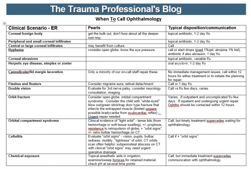
When To Call: Ophthalmology

Here’s another in my series of “When To Call” pieces. We sometimes overuse our consultants and call then at inappropriate times. So what if we diagnose an injury in their area of expertise at 2 am? Does it need attention or an operation before morning? If not, why call at that ungodly hour?

Let’s use our consultants wisely! I’ve listed most of the common eye diagnoses that trauma professionals will encounter. There is also an indication of what you need to do, and exactly when to call your consultant.

Unfortunately, this one won’t fit on a 3×5 index card that you can keep in your pocket. I’ve included a printable pdf file, as well as the original Microsoft Word file in case you want to make a few modifications to suit your own hospital.

Enjoy!

When to call Ophthalmology reference card (pdf)

When to call Ophthalmology MS Wordfile (docx)

Management Of Blunt Carotid / Vertebral Injury

Yesterday I reviewed the most commonly used grading system for blunt carotid / vertebral injury (BCVI). Today, I’ll describe the usual management of these injuries, by grade. Unfortunately, there is a paucity of definitive literature to guide us because these injuries are rare. So here are our best guesses to date.

There are basically three modalities at our disposal for managing BCVI: antithrombotic medication (heparin and/or antiplatelet agents), surgery, and therapeutic angiographic procedures. The choice of therapy is usually based on surgical accessibility and patient safety for anticoagulation. We do know that a number of studies have shown a decrease in stroke events in patients who are heparinized. Unfortunately, this is not always possible due to associated injuries. Antiplatelet agents are usually tolerated after acute trauma, especially low-dose aspirin. Several studies have shown little difference in outcomes in patients receiving heparin vs aspirin/clopidogrel for BCVI.

So what to do? Here are some broad guidelines:

  • Grade I (intimal flap). Heparin or antiplatelet agents should be given. If heparin can be safely administered, it may be preferable in patients who will need other surgical procedures since it can be rapidly reversed just by stopping the infusion. These lesions generally heal completely, so a followup CT angiogram should be scheduled in 1-2 weeks. Medication can be stopped when the lesion heals.
  • Grade II (flap/dissection/hematoma). These injuries are more likely to progress, so heparin is preferred if it can be safely given. Stenting should be considered, especially if the lesion progresses. Long-term anti-platelet medication may be required.
  • Grade III (pseudoaneurysm). Initial heparin therapy is preferred unless contraindicated. Stable pseudoaneurysms should be followed with CTA every 6 months. If the lesion enlarges, then surgical repair should be carried out in accessible injuries, or stenting in inaccessible ones.
  • Grade IV (occlusion). Heparin therapy should be initiated unless contraindicated. Patients who do not suffer a catastrophic stroke may do well with followup antithrombotic therapy. Endovascular treatment does not appear to be helpful.
  • Grade V (transection with extravasation). This lesion is frequently fatal, and the bleeding must be addressed using the best available technique. For lesions that are surgically accessible, the patient should undergo the appropriate vascular procedure. Inaccessible injuries should undergo angiographic treatment, and may require embolization to control bleeding without regard for the possibility of stroke.

References:

  • Scott WW, Sharp S, Figueroa SA, et al. Clinical and radiographic outcomes following traumatic Grade 1 and 2 carotid artery injuries: a 10-year retrospective analysis from a Level I trauma center. J Neurosurg 122:1196, 2015.
  • Scott WW, Sharp S, Figueroa SA, et al. Clinical and radiographic outcomes following traumatic Grade 3 and 4 carotid artery injuries: a 10-year retrospective analysis from a Level 1 trauma center. J Neurosurg 122:610, 2015.
  • Scott WW, Sharp S, Figueroa SA, et al. Clinical and radiological outcomes following traumatic Grade 1 and 2 vertebral artery injuries: a 10-year retrospective analysis from a Level 1 trauma center. J Neurosurg 121:450, 2015.
  • Scott WW, Sharp S, Figueroa SA, et al. Clinical and radiological outcomes following traumatic Grade 3 and 4 vertebral artery injuries: a 10-year retrospective analysis from a Level I trauma center. The Parkland Carotid and Vertebral Artery Injury Survey. J Neurosurg 122:1202, 2015.


What Does Blunt Carotid / Vertebral Injury Look Like?

In my last two posts, I reviewed who is at risk for blunt carotid / vertebral injury (BCVI) and how to find it. But what does it actually look like, and how is it classified?

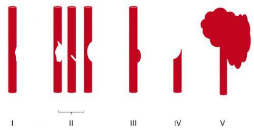
A seminal paper from Denver Health (aka Denver General Hospital) in 1999 proposed the most commonly used grading system for BCVI. This Denver scale should not be confused with the Denver criteria that predict risk for BCVI. Here’s a nice graphic that explains the classifications:

Grade I: A mild intimal irregularity is seen. Note the abnormal  narrowed area, representing a small intimal injury, possibly with a small amount of clot.

Grade II: This grade has several presentations. There may be a intraluminal thrombosis/hematoma with (left) or without (right) an intimal flap, or a flap alone (center)

Grade III: There is a full-thickness injury to the vessel with a contained extraluminal extravasation (pseudoaneurysm)

Grade IV: The vessel is completely occluded by flap or thrombus

Grade V: The artery is transected and freely extravasating

In the next post, I’ll finish off with a summary of the treatments for these injuries.

Reference: Blunt carotid arterial injuries: implications of a new grading scale. J Trauma. 47(5): 845-53, 1999.

Who Is At Risk For Blunt Cerebrovascular Injury?

In my last post, I wrote about proper screening for blunt cerebrovascular injury (BCVI). But, as you know, it’s important to screen only when there is a significant risk of the injury being present. Screening using the shotgun approach (screen everyone for everything) yields enough false positive results to present potential danger to your patient.

A variety of authors on this topic have promoted a number of high risk criteria to trigger a screening test. Most make sense, and are related to the anatomy of the vessels in question. The carotid arteries are relatively unprotected, although a bit deep, as they course up the neck. Thus, it is possible to damage them when they suffer a direct and significantly hard blow. Once they enter the skull, they are better protected. However, fractures through key areas of the skull base and face can injure the vessels, even in these protected locations.

The vertebral arteries are deep and relatively protected as they course through the vertebral foramina. However, if the vertebrae are fractured or subluxed, vessel injury can occur.

Finally, and as always, the physical exam is important. If there are unexpected neurologic changes that can’t be explained by other injuries, or there are indications of deep vascular injury, BCVI needs to be considered.

Here is my list of indications to screen for BCVI:

  • Neurologic abnormality not explained by diagnosed injury
  • Arterial epistaxis†
  • Seat belt sign on neck†
  • GCS < 8 (this is the most commonly forgotten one)
  • Petrous bone fracture
  • C‐spine fracture (C1‐C3) or subluxation at any level†
  • Fracture through foramen transversum†
  • LeFort II or III fractures

Bottom line: Be on the lookout for any of the criteria listed above in your trauma patient. If you find one during your initial evaluation, be sure to order a CT angiogram of the neck. And keep an eye out while scanning the head and cervical spine. If any of the other radiographic indications become apparent, add on the CT angiogram at that point.