A Blood Test For TBI? Part 2

Yesterday, I wrote about one blood biomarker, GFAP, and its possible application in detecting traumatic brain injury (TBI). Today, I’ll discuss a complementary marker called UCH-L1.

Fewer studies have been done looking at the utility of UCH-L1 in detecting TBI than of GFAP.  A review article published last month pooled existing literature to get a sense of how good this biomarker really is. It also examined the risk of bias due to the small numbers of studies involved.

Here are the factoids:

  • Only 38 abstracts were eligible, but full text was available for analysis in only 13 (meaning it was only an abstract and never passed muster for publication). The authors of the published studies were contacted for additional information, which is an interesting (and helpful) practice.
  • Of all of those, only 4 were selected for meta-analysis! This significantly limited the value of the analysis.
  • Serum UCH-L1 has a high accuracy in predicting CT findings in mild to moderate TBI, but there is a high risk of bias affecting this result
  • Plasma UCH-L1 has a moderate accuracy predicting CT findings across all GCS levels, with a low risk of bias
  • Pooling all studies, this is high accuracy in predicting CT findings in patients with TBI across all GCS levels, but there is a high risk of bias affecting the results

Bottom line: UCH-L1 show promise as a predictor of CT findings in patients with TBI. However, the research papers were few and far between, and the possibility of bias was high. What does this mean? That using this test alone is better than a coin toss, but not good enough to change our practice in ordering CT scans in head injured patients. More well-designed studies are needed tell us whether this new (and undoubtedly expensive) test is worth the trouble.

Tomorrow, I’ll discuss a blood test incorporating both UCH-L1 and GFAP that was recently approved by the FDA.

Reference: The diagnostic values of UCH-L1 in traumatic brain injury: A meta-analysis. Brain Inj 32(1):1-17, 2018.

A Blood Test For TBI? Part 1

Traumatic brain injury (TBI) is an extremely common problem encountered by trauma professionals. Diagnostic and management pathways are fairly well-defined, and rely mainly on physical examination, as well as CT imaging in select cases.

In recent years, work has been done to identify markers of brain injury in the blood. The theory is that the injured brain may release substances that can be assayed with a simple blood test. The presence of these blood markers could then influence our use of CT for diagnosis, decision to admit or send home, and possibly help identify patients likely to develop post-concussive symptoms.

Two particular biomarkers are being evaluated: UCH-L1 and GFAP. A recently published review examined the current status of GFAP in diagnosis of head injury.

Here are the factoids:

  • A total of 27 pertinent research papers were identified for review, and 24 of 27 demonstrated a positive association between GFAP levels and TBI
  • GFAP prediction of intracranial pathology by CT scan was good to excellent
  • GFAP appeared to be able to discriminate between mass lesions and diffuse injury
  • There was considerable variability in the average GFAP values. This means that the cutoff value that predicts significant injury is not yet clear.
  • The number of pediatric studies reviewed was low, so the results may not be generalizable to children
  • GFAP may be elevated in patients with orthopedic injuries, and this was not well controlled for in the studies reviewed. It is unclear whether GFAP can be used in patients with fractures.

Bottom line: GFAP looks promising as a marker for detecting significant TBI in some trauma patients. 

Tomorrow, I’ll take a look at the other biomarker, UCH-L1, and the following day I’ll discuss the recent FDA approval of an assay for both of these by a US company, Banyan Biomarkers.

Reference: A systematic review of the usefulness of glial fibrillary acidic protein for predicting intracranial lesions following head trauma. Frontiers in Neurology 8(652):1-16.

I have no financial interest in Banyan Biomarkers.

How To Evaluate A Stab To The Diaphragm – Part 2

Yesterday I gave a little perspective on the use of CT in assessing the diaphragm after penetrating injury. Today, I’ll break it down into some practical steps you can follow the next time you see one.

Step 1. Stable or unstable? If your patient arrives with unstable vital signs, and there is no other source but the abdomen, the answer is simple. Go to the OR for a laparotomy. Period. They are exsanguinating and the hemorrhage needs to be stopped.

Step 2. Mark the sites of penetration and take a chest x-ray. This will let you evaluate the potential trajectory of the object, and will give you your first glimpse of the diaphragm.

Step 3. Examine the abdomen. Actually, you should be doing this at the same time you are setting up for Step 2. If your patient has peritoneal signs, no further evaluation is needed. Just go to the OR for laparotomy. Look at the chest x-ray once you get there.

Step 4. Right side? If your appreciation of the path of penetration involves just the liver, take the patient to CT for evaluation of chest, abdomen, and pelvis. You need to see all three of these areas to assess for blood and fluid in both body cavities. After the study, if you still think the injury is limited to the liver, admit the patient for observation.

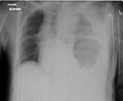
Step 5. Left side? Look at that chest x-ray again. If there are any irregularities at all, strongly consider going to the OR and starting with diagnostic laparoscopy. These irregularities can be glaring, like in the x-ray above. But they can be subtle, like some haziness above the diaphragm or small hemothorax. Obviously, if the injury is as clear as on the x-ray above, just open the abdomen. But if in doubt, start small. And remember my advice on “lunchothorax.”

Step 6. Admit and observe. Check the abdomen periodically, and repeat the chest x-ray daily. If anything changes, consider diagnostic laparoscopy. As a general rule, I don’t keep patients NPO “just in case.” Most will pass this test, and I don’t see a reason to starve my patients for the low likelihood they need to go to the OR.

Step 7. Make sure your patient gets a follow up evaluation. See them in your outpatient clinic, get a final chest x-ray and abdominal exam before you completely clear them.

How To Evaluate A Stab To The Diaphragm – Part 1

Penetrating injury to the diaphragm, and specifically stab wounds, have been notoriously hard to diagnose since just about forever. Way back in the day (before CT), we tried all kinds of interesting things to help figure out if the patient had a real injury. Of course, we could just go to the OR and lap the patient (laparoscopy did not exist then). But the negative lap rate was significant, so we tried a host of less invasive techniques.

Remember diagnostic peritoneal lavage? Yeah, we tried that. The problem was that the threshold for red cells per cubic mm was not well defined. Some would supplement this technique with a chest tube to see if lavage fluid would drain out. And one paper described instilling nuclear medicine tracer into the abdomen and sitting the patient under a gamma camera for a few hours to see if any ended up in the chest. Groan!

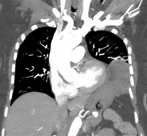
We thought that CT would save us. Unfortunately, resolution was terrible in the early years. If you could actually see the injury on CT, it was probably because a large piece of stomach or colon had already fallen through it. But as detectors multiplied and resolution improved, we could begin to see some smaller defects. But we still missed a few. And the problem is that left-sided diaphragmatic holes slowly enlarge over time (years), until the stomach or colon falls through it. (See below)

A group of radiologists and surgeons in a Turkish trauma hospital recently published a modest series of patients with left-sided diaphragm injuries evaluated by CT. They looked at about 5 years of their experience in a group of patient who were at risk for the injury due to a thoraco-abdominal stab wound. Unstable patients were immediately taken to OR. All of the remaining patients underwent an initial CT scan, followed by diagnostic laparoscopy after 48 hours if they remained symptom free.

Here are the factoids:

  • A total of 43 stable patients with a left thoraco-abdominal stab were evaluated
  • 30 patients had a normal CT, and 13 had the appearance of an injury
  • Of those who were CT positive, only 9 of 13 (69%) actually had the injury at operation
  • Two of the 30 (7%) who were CT negative were found to have a diaphragm injury during followup laparoscopy
  • So in the author’s hands, there was 82% sensitivity, 88% specificity, a positive predictive value of only  69%, and a negative predictive value of 93%

Bottom line: The authors somehow looked at the numbers and concluded that CT is valuable for detecting left diaphragm injury. Huh? They missed 7% of injuries, only finding them later at laparoscopy. And they had a 31% negative laparotomy rate. 

Now, it could be that the authors were using crappy equipment. Nowhere in their paper do they state how many detectors, or what technique was used. Since it took place over a 5 year period, it is quite possible that the earlier years of the study used equipment now considered to be out of date, or that there was no standardized technique.

CT may not yet be ready for prime time. But it can be a valuable tool. Tune in tomorrow for some tips on how and when to look for this insidious injury.

Reference: Evaluation of diaphragm in penetrating left thoracoabdominal
stab injuries: The role of multislice computed tomography. Injury 46:1734-1737, 2015.

What Is: Lunchothorax?

Here’s an operative tip for trauma professionals who find themselves in the OR. Heard of “lunchothorax?” I’m sure most of you haven’t. The term originated in a 1993 paper on the history of thoracoscopic surgery. It really hasn’t been written about in the context of trauma surgery, though.

Lunchothorax is an empyema caused by pleural contamination in patients with concomitant diaphragm and hollow viscus injury. This most commonly occurs with penetrating injuries to the left upper quadrant and/or left lower back. The two penetrations tend to be in close proximity (diaphragm + stomach), but may occasionally be further away (diaphragm + colon).

One of the earlier papers describing the correlation of gastric injury and empyema was written by one of my mentors, John Weigelt. Although gastric repair is usually simple and heals well, his group did note a few severe complications. Of 243 patients with this injury, 15 developed ones that were considered severe, and 10 of those were empyema! What gives?

It turns out that the combination of gastric contents and pleural space is not a good one. It’s not really clear why this is. Is it bacterial? The acid? Undigested food? I’ve seen cases with what I would consider minimal contamination go on to develop a nasty empyema. This is also borne out in a National Trauma Databank review from 2009. It looked at complications in patients with a diaphragm injury and found that a gastric injury increased the probability of empyema by 3x. Interestingly, there was no increased risk of empyema with a concomitant colon injury.

Bottom line: Lunchothorax, or empyema after even minimal contamination from a hollow viscus, is a dreaded complication of thoraco-abdominal penetrating injury. Any time the stomach and diaphragm are violated, I recommend thoroughly irrigating the chest. It’s probably a good idea for concomitant colon injury as well, but there’s less literature support.

This can be done through the diaphragm injury if it is large enough, or through a chest tube inserted separately. Most of the time, you’ll be placing the chest tube anyway because the pleural space has been violated via the abdomen. In either case, copious lavage with saline is recommended to clear all particulate material, with a few extra liters just for good measure. There’s no data on use of antibiotics, but standard perioperative coverage for the abdominal injuries should be sufficient if the lavage was properly performed.

References:

  • The history of thoracoscopic surgery. Ann Thoracic Surg 56(3):610-614, 1993.
  • Penetrating injuries to the stomach. SGO 172(4):298-302, 1991.
  • Risk factors for empyema after diaphragmatic injury: results of a National Trauma Databank analysis. J Trauma 66(6):1672-1676, 2009.