Post-Embolization Syndrome In Trauma

A reader requested that I write about post-embolization syndrome. Not being an oncologist or oncologic surgeon, I honestly had never heard about this before, let alone in trauma care. So I figured I would read up and share. And fortunately it was easy; there’s all of one paper about it in the trauma literature.

Post-embolization syndrome is a constellation of symptoms including pain, fever, nausea, and ileus that occurs after angio-embolization of the liver or spleen. There are reports that it is a common occurrence (60-80%) in patients being treated for cancer, and there are a few papers describing it in patients with splenic aneurysm. But only one for trauma.

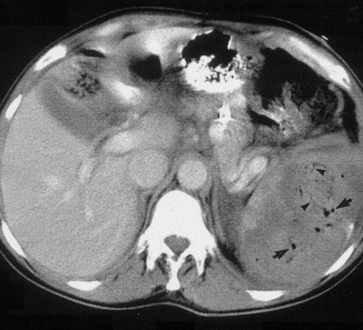
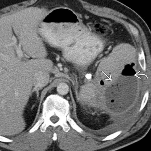
Children’s Hospital of Boston / Harvard Medical School retrospectively reviewed 12 years of their pediatric  trauma registry data. For every child with a spleen injury who underwent angio-embolization, they matched four others with the same grade of injury who did not. A total of 448 children with blunt splenic injury were identified, and (thankfully) only 11 underwent angio-embolization. Nine had ongoing bleeding despite resuscitation, and two had developed splenic pseudoaneursyms.

Here are the factoids:

  • More of the children who underwent embolization had extravasation seen initially and required more blood products.  They also had longer ICU (3 vs 1 day) and hospital stays (8 vs 5 days). Not surprising, as that is why they had the procedure.
  • 90% of embolized kids had an ileus vs 2% of those not embolized, and they took longer to resume regular diet (5 vs 2 days)
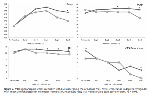
  • Respiratory rate and blood pressure were higher on days 3 and 4 in the embolized group, as was the temperature on day 5 (? see below)
  • Pain was higher on day 5 in the embolized group (? see below again)

Bottom line: Sorry, but I’m not convinced. Yes, I have observed increased pain and temperature elevations in patients who have been embolized. Some have also had an ileus, but it’s difficult to say if that’s from the procedure or other injuries. And this very small series just doesn’t have enough power to convince me of any clinically significant differences in injured children.

Look at the results above. “Significant” differences were only identified on a few select days, but not on the same days across charts. And although the authors may have demonstrated statistical differences, are they clinically relevant? Is a respiratory rate of 22 different from 18? A temp of 37.8 vs 37.2? I don’t think so. And length of stay does not reveal anything because the time in the ICU or hospital is completely dependent on the whims of the surgeon.

I agree that post-embolization syndrome exists in cancer patients. But the findings in trauma patients are too nondescript. They just don’t stand out well enough on their own for me to consider them a real syndrome. As a trauma professional, be aware that your patient probably will experience more pain over the affected organ for a few days, and they will be slow to resume their diet. But other than supportive care and patience, nothing special need be done.

Related posts:

Reference: Transarterial embolization in children with blunt splenic injury
results in postembolization syndrome: A matched
case-control study. J Trauma 73(6):1558-1563, 2012.

Incidental Finding: Gas In The Spleen After Embolization

Most solid organ injury practice guidelines include angioembolization in part of the pathway. But very few require re-imaging at any point to see how the liver or spleen are coming along.

But every once in a while another condition arises, or symptoms worsen unexpectedly, causing us to get another CT scan that includes the abdomen and pelvis. And sometimes we see things that we wouldn’t normally see, like air bubbles in the organ that was embolized.

So what is okay, and what requires some kind of intervention? Our friends at ShockTrauma in Baltimore looked at this in 2001 and can provide some pretty good guidance. They reviewed patients who underwent CT scan both before and after embolization over about 2.5 years. They performed the post-embolization scans for specific indications like fevers, elevated WBC count (!), increasing abdominal pain, or an episode of hypotension. A total of 53 patients were studied.

Here are the factoids:

  • 24 patients underwent embolization of the main splenic artery, 22 had selective embolization of part of the spleen, and 7 had both
  • Splenic infarcts occurred in 63% of patients with main artery embolization, but were large (> 50% of the parenchyma) in only 20% of those
  • Infarcts occurred in 100% of selective embolizations, but were small (< 50%) in 93% of cases
  • Infarcts occurred in 71% of patients with both main and selective embolization, and most were small (80%)
  • Seven (13%) patients developed gas bubbles in the spleen, and was usually present for 1-7 days before disappearing
  • One patient developed increasing gas with pneumoperitoneum and underwent splenectomy for a splenectomy for abscess

This picture that shows tiny bubbles in the spleen parenchyma that represent “normal” gas after embolization:

And the following one shows an air/fluid collection in the spleen that indicates an abscess:

Bottom line: Tiny bubbles in the spleen (and probably the liver) occur normally after angioembolization. They usually develop within an area of infarction, and most are benign. It is possible for them to evolve into a splenic abscess, but unlikely. Many embolization patients develop fevers at some point, and most have an elevated WBC count. So in most cases, you can ignore this incidental finding, as long as your patient has mild symptoms.

However, if the patient develops high fevers, very elevated WBC (> 25K), increasing abdominal or flank pain, and the spleen develops an air/fluid level, an abscess is forming. Despite what your radiologist might suggest, catheter drainage is not a good idea. The tubes are too small to remove the slurry that is generally found within the abscess. A trip to the OR is the only effective treatment, and splenectomy is generally the only option.

Related posts:

Reference: CT Findings after Embolization for Blunt Splenic Trauma. J Vasc Intervent Radiol 12(2):209-214, 2001.

Everything You Wanted To Know About: Cranial Bone Flaps

Patients with severe TBI frequently undergo surgical procedures to remove clot or decompress the brain. Most of the time, they undergo a craniotomy, in which a bone flap is raised temporarily and then replaced at the end of the procedure.

But in decompressive surgery, the bone flap cannot be replaced because doing so may increase intracranial pressure. What to do with it?

There are four options:

  1. The piece of bone can buried in the subcutaneous tissue of the abdominal wall. The advantage is that it can’t get lost. Cosmetically, it looks odd, but so does having a bone flap missing from the side of your head. And this technique can’t be used as easily if the patient has had prior abdominal surgery.

2. Some centers have buried the flap in the subgaleal tissues of the scalp on the opposite side of the skull. The few papers on this technique demonstrated a low infection rate. The advantage is that only one surgical field is necessary at the time the flap is replaced. However, the cosmetic disadvantage before the flap is replaced is much more pronounced.

3. Most commonly, the flap is frozen and “banked” for later replacement. There are reports of some mineral loss from the flap after replacement, and occasional infection. And occasionally the entire piece is misplaced. Another disadvantage is that if the patient moves away or presents to another hospital for flap replacement, the logistics of transferring a frozen piece of bone are very challenging.

4. Some centers just throw the bone flap away. This necessitates replacing it with some other material like metal or plastic. This tends to be more complicated and expensive, since the replacement needs to be sculpted to fit the existing gap.

So which flap management technique is best? Unfortunately, we don’t know yet, and probably never will. Your neurosurgeons will have their favorite technique, and that will ultimately be the option of choice.

Reference: Bone flap management in neurosurgery. Rev Neuroscience 17(2):133-137, 2009.

The  March Trauma MedEd Newsletter Is Available!

This month’s newsletter contains a veritable potpourri of topics that I found interesting. I hope you do, as well. Here are the topics covered:

  • Prehospital lactate levels: useful or not?
  • Does aspirin for DVT prophylaxis work?
  • The nuts and bolts of Duplex ultrasound
  • An 11 year experience with endovascular repair of aortic injuries
  • Use of MRI after CT scanning of the cervical spine

Subscribers received this issue last week. Subscribe now and be sure to get the next issue early.  So sign up for early delivery now by clicking here!

Click here to download the current issue

Pick up back issues here!


Pet Peeve: “High Index of Suspicion”

How often have you heard this phrase in a talk or seen it in a print article:

“Maintain a high index of suspicion”

What does this mean??? It’s been popping up in papers and textbooks for at least the last 20 years. And to me, it’s meaningless. You try to figure out that sentence!

An index is a number, usually mathematically derived in some way. Yet whenever I see or hear this phrase, it doesn’t really apply to anything that is quantifiable. What the author is really referring to is “a high level of suspicion”, not an index.

This term has become a catch-all to caution the reader or listener to think about a (usually) less common diagnostic possibility. As trauma professionals, we are advised to do this about so many things, it really has become sad and meaningless.

Bottom line: Don’t use this phrase in your presentations or your writing. It’s stupid. And feel free to chide any of your colleagues who do. Give us some concrete tips so we don’t miss the diagnosis!

Reference: High index of suspicion. Ann Thoracic Surg 64:291-292, 1997.