CT Scan Image Settings Simplified

Ever wonder what is going on when you drag your mouse across a CT image, or when you change the “window” settings of an image from lung to abdomen? It all has to do with the way CT generated xray information is displayed, and how your eyes and brain perceive it.

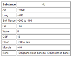
Let’s get down to basics. The first thing needed is to understand the concept of radiodensity. The CT scanner uses a set of software algorithms to determine the amount of x-radiation absorbed by every element in a plane of tissue. Each of these elements is represented by a pixel on the video display, and the density (amount of x-radiation absorbed) is measured in Hounsfield units. This scale was developed by Sir Godfrey Hounsfield, who set the radiodensity of water at 0, and air at -1000. The scale extends in the positive direction to about +4000, which represents very dense metals. See the table for the density of common substances on CT.

When you view a CT scan on a video display, two important numbers are displayed on screen. The first is the window width (W), which describes the range of Hounsfield units displayed. The maximum window width possible is usually about 2000, but our eyes are not capable of seeing this many shades. Actually, we can really only distinguish about 16 shades of gray. So the window width is divided by 16, and each group of Hounsfield values is converted to one of 16 shades of gray. The lowest Hounsfield numbers in the window range are shown as black, and the highest are white.

The second important number is the window level (L). This is the Hounsfield number in the center of the window width. So let’s look at some typical examples of W/L settings.

The abdomen contains mostly soft tissue, which is just a little denser than water. So most of the abdominal contents have Hounsfield values from 0 to 100 or so. A typical abdominal scan W/L setting is 350/50. This means that a total range of 350 different densities are displayed, centered on a density of 50 Hounsfield units ( range is -125 to 225 HU). Each difference of 22 HU will show up as a different shade of gray. So this narrow window allows us to distinguish relatively subtle differences in density.

The chest cavities are primarily air-filled, and the lungs are very low density. So it makes sense that a typical lung W/L setting is 1500/-500. The window ranges from -1250 to +250 HU, and a wider range of 94 HU represents one shade of gray. This is typical of body regions with a wider range of densities.

Finally, bone windows are usually 2000/250. This window is centered above the usual tissue densities, and is very wide so that it shows a wide range of densities in only 16 shades of gray. Thus, the contrast appears very low.

On most displays, the window width increases as you drag the mouse to the right. This increases the range of densities in a shade of gray, thus decreasing the overall amount of contrast in the image. Dragging the mouse down decreases the window level, moving it toward the air end of the spectrum. This allows you to center your window on the type of tissue you are interested in viewing and adjust your ability to distinguish objects with a lot or only a little contrast (see table above).

I apologize to my radiology colleagues in advance for this simplistic explanation. Trauma professionals have minimal exposure (pun intended) to the physics and details of radiographic imaging. We are much more interested in effectively using this technology to save our patients’ lives.

Acute Ethanol Intoxication And The Banana Bag

It’s one of those time honored treatments that most hospital-based providers are familiar with. The banana bag, reserved for intoxicated patients presenting to the ED or admitted to the hospital. They’ve been around so long, we just take them for granted. But like most things that have become dogmatic, they are due to be questioned from time to time.

A banana bag is a proprietary mix of “good” stuff, including electrolytes and vitamins, especially thiamine and magnesium. The exact content varies from hospital to hospital. Thiamine and other B vitamins give the resulting solution the characteristic color, hence the term “banana.”

Does it actually do good things like ward off Wernicke’s encephalopathy and megaloblastic anemia? A paper from Jacobi Medical Center in the Bronx prospectively evaluated a series of intoxicated people entering their ED. They drew vitamin B12, folate, and thiamine levels to see if they were deficient enough to even need vitamin supplementation.

Interesting findings:

  • These folks (only 77 patients) were very drunk! Average BAC was 280mg/dL.
  • Vitamin B12 and folate levels were not critically low in any patient
  • Thiamine was low in 15% of patients, but none had clinical evidence of a deficiency
  • Later review of prior visits revealed that some patients with low levels had received a previous banana bag within 1 month. Did it do any good?

Bottom line: Most of our intoxicated patients are not vitamin deficient, and don’t need supplementation. The real kicker is that  we almost never really try to find out if the patient might be a chronic abuser and potentially at risk. We just hang the bag. Remember, everything we do in medicine has a potential downside. And if the patient really doesn’t need a banana bag in the first place, there is no benefit to balance that risk. The next time you ask for that little yellow bag, think again!

Reference: Vitamin deficiencies in acutely intoxicated patients in the ED. Am J Emerg Med 26(7):729-795, 2008.

Are We Transporting Our Patients In The Correct Position? Part 2

[Note to prehospital providers: please comment below or email with your experience using this position.]

In my last post, I discussed the only paper I could find on the lateral trauma position (LTP). It was a survey that was taken 5 years after implementation of this transport position in Norway. Is there anything else out there that may help give us guidance on proper positioning during transport?

Just this month, a paper was published that tries to look at this issue from a different viewpoint. Since we can’t really show that the LTP is good or prove that it is truly safe, can we at least demonstrate that supine positioning might be bad?

 A very diverse group of researchers in Norway performed a systematic literature review and meta-analysis of everything they could find published on supine positioning and airway patency in unconscious trauma patients, especially when compared to lateral positioning. This was carried out from the beginning of time, or 1959 in this case.

See if you can follow their progress:

  • There weren’t really any good studies using this global search, so they broadened it to include trauma patients with decreased level of consciousness.
  • Oops! There weren’t any studies using this broader definition, either.
  • The authors wanted to use morbidity and mortality as their outcomes. But, there weren’t any good studies for this either so the decided to use indirect outcomes such as hypoxia, hypercapnea, hypoventilation, work of breathing, and a bunch of other stuff.
  • Oops again! There weren’t any studies reporting these indirect outcomes. 
  • But when these two indirect searches were combined, a number of papers (20) were identified that were used for a meta-analysis
  • A number of these papers showed soft results (language like ”indication of”, “small difference”). The only significant results were found in patients with known obstructive sleep apnea.

Bottom line: The use of the lateral trauma position is an intriguing concept, and has been used successfully in Norway for about 10 years. Intuitively, it makes sense, especially in obese patients or those with known obstructive sleep apnea. Unfortunately, this paper approached the questions asked kind of backwards, in my opinion.

I believe that LTP has a place in prehospital care, but that there will be significant barriers to adoption in most countries. In order to overcome these hurdles, clear protocols and positioning instructions will need to be developed, as well as specific indications. And it wouldn’t hurt to do a few good studies along the way. The Norwegians have helped us with the ethics questions, as it is the standard of care in that country. So write your local IRB and get busy!

Related post:

Reference: Is the supine position associated with loss of airway patency in unconscious trauma patients? A systematic review and meta-analysis. Scand J Trauma Resusc Emerg Med 23:50, July 1, 2015. 

Are We Transporting Our Patients In The Correct Position? Part 1

Here’s a kick in the head, unless you are one of my Norwegian readers.

In the US and many (most) other countries, trauma patients are routinely transported strapped down in the supine position. It’s tradition. It’s easy. It gives prehospital providers pretty good access for whatever they need to do.

But it is right? Some patients, particularly those who have a diminished level of consciousness or severe obstructive sleep apnea, or both, may not do well in that position. In 2005, Norwegian Emergency Medical Services (NEMS) introduced the use of the lateral trauma position (LTP) across the country. Here is how it looks:

Five years later, a group from several hospitals across Norway conducted a survey of all ground and air EMS providers in the country. A few factoids:

  • This one year survey included 202 of 206 ground EMS stations and 23 of 24 air EMS stations. Questionnaire response rate was about 50%
  • Of supervisors at ground EMS units, 75% said that they had implemented LTP
  • 67% of ground units had written policies for use and 73% had provided training
  • Individual ground provider opinions were a bit different. 89% were familiar with LTP, but only 58% actually used it.
  • Training seemed to be the key. Of ground providers given training, 86% were confident in using LTP, but of those not given training, only 58% were.
  • Only 53% of air services used LTP, and only one had a protocol.

Here are the instructions on how to do it:

Bottom line: Interesting concept. Unfortunately there is little (or no) objective data to help us. The main thing available now is a 10 year experience with the lateral trauma position in Norway, and I have not seen any analyses of it. In my next post, I’ll review a meta-analysis published this year that does try to compare LTP vs supine positioning.

Related posts:

Reference: The lateral trauma position: what do we know about it and how do we use it? A cross-sectional survey of all Norwegian emergency medical services. Scand J Trauma 19:45, 2011.

What Is: An “Egg Timer Injury”?

Most patients with major traumatic injuries are handled in a very systematic way by both EMS and trauma centers. We have routines and protocols designed to provide rapid, quality care to these individuals. But over the years, I’ve begun to appreciate the fact that there is a very small subset of these patients who are different.

I term these patients as having an “egg timer injury”. These are patients who have only a certain number of minutes to live. This fact requires us to change the usual way we do things in order to save their lives or limbs. The usual routine may be too slow.

And unfortunately, no one can tell us exactly how many minutes are left on the timer. We only know that it’s ticking. Here are some examples of such  injuries:

  • Pericardial tamponade
  • Penetrating injury to the torso with profound hypotension
  • Orbital compartment syndrome

In each case, speed is of the essence. What can we do to decrease the time to definitive intervention? For prehospital providers, you may need to bypass a closer hospital that might not have the necessary resources at a particular time of day. Once at the hospital, the patient may need to bypass the emergency department and proceed straight to the OR. Or you may need to do a lateral canthotomy yourself, rather than waiting for an ophthalmologist to drive in only to have the patient lose their vision because of the  delay.

Bottom line: Remember that protocols are not necessarily etched in stone. They will cover 99.9% of cases you see. But that remaining 0.1%, the patients with the “egg timer injury”, will require you to think through what you know about the patient at the time, and make decisions about their care that may have a huge outcome on their life or livelihood. And as always, if you find that you must do things differently in the best interest of your patient, be sure to document what you knew and your thought processes thoroughly so you explain and/or justify your decision-making when you are invariably asked.