All posts by The Trauma Pro

Management Of Blunt Carotid / Vertebral Injury

Yesterday I reviewed the most commonly used grading system for blunt carotid / vertebral injury (BCVI). Today, I’ll describe the usual management of these injuries, by grade. Unfortunately, there is a paucity of definitive literature to guide us because these injuries are rare. So here are our best guesses to date.

There are basically three modalities at our disposal for managing BCVI: antithrombotic medication (heparin and/or antiplatelet agents), surgery, and therapeutic angiographic procedures. The choice of therapy is usually based on surgical accessibility and patient safety for anticoagulation. We do know that a number of studies have shown a decrease in stroke events in patients who are heparinized. Unfortunately, this is not always possible due to associated injuries. Antiplatelet agents are usually tolerated after acute trauma, especially low-dose aspirin. Several studies have shown little difference in outcomes in patients receiving heparin vs aspirin/clopidogrel for BCVI.

So what to do? Here are some broad guidelines:

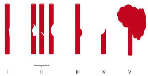
  • Grade I (intimal flap). Heparin or antiplatelet agents should be given. If heparin can be safely administered, it may be preferable in patients who will need other surgical procedures since it can be rapidly reversed just by stopping the infusion. These lesions generally heal completely, so a followup CT angiogram should be scheduled in 1-2 weeks. Medication can be stopped when the lesion heals.
  • Grade II (flap/dissection/hematoma). These injuries are more likely to progress, so heparin is preferred if it can be safely given. Stenting should be considered, especially if the lesion progresses. Long-term anti-platelet medication may be required.
  • Grade III (pseudoaneurysm). Initial heparin therapy is preferred unless contraindicated. Stable pseudoaneurysms should be followed with CTA every 6 months. If the lesion enlarges, then surgical repair should be carried out in accessible injuries, or stenting in inaccessible ones.
  • Grade IV (occlusion). Heparin therapy should be initiated unless contraindicated. Patients who do not suffer a catastrophic stroke may do well with followup antithrombotic therapy. Endovascular treatment does not appear to be helpful.
  • Grade V (transection with extravasation). This lesion is frequently fatal, and the bleeding must be addressed using the best available technique. For lesions that are surgically accessible, the patient should undergo the appropriate vascular procedure. Inaccessible injuries should undergo angiographic treatment, and may require embolization to control bleeding without regard for the possibility of stroke.

References:

  • Scott WW, Sharp S, Figueroa SA, et al. Clinical and radiographic outcomes following traumatic Grade 1 and 2 carotid artery injuries: a 10-year retrospective analysis from a Level I trauma center. J Neurosurg 122:1196, 2015.
  • Scott WW, Sharp S, Figueroa SA, et al. Clinical and radiographic outcomes following traumatic Grade 3 and 4 carotid artery injuries: a 10-year retrospective analysis from a Level 1 trauma center. J Neurosurg 122:610, 2015.
  • Scott WW, Sharp S, Figueroa SA, et al. Clinical and radiological outcomes following traumatic Grade 1 and 2 vertebral artery injuries: a 10-year retrospective analysis from a Level 1 trauma center. J Neurosurg 121:450, 2015.
  • Scott WW, Sharp S, Figueroa SA, et al. Clinical and radiological outcomes following traumatic Grade 3 and 4 vertebral artery injuries: a 10-year retrospective analysis from a Level I trauma center. The Parkland Carotid and Vertebral Artery Injury Survey. J Neurosurg 122:1202, 2015.


What Does Blunt Carotid / Vertebral Injury Look Like?

In my last two posts, I reviewed who is at risk for blunt carotid / vertebral injury (BCVI) and how to find it. But what does it actually look like, and how is it classified?

A seminal paper from Denver Health (aka Denver General Hospital) in 1999 proposed the most commonly used grading system for BCVI. This Denver scale should not be confused with the Denver criteria that predict risk for BCVI. Here’s a nice graphic that explains the classifications:

Grade I: A mild intimal irregularity is seen. Note the abnormal  narrowed area, representing a small intimal injury, possibly with a small amount of clot.

Grade II: This grade has several presentations. There may be a intraluminal thrombosis/hematoma with (left) or without (right) an intimal flap, or a flap alone (center)

Grade III: There is a full-thickness injury to the vessel with a contained extraluminal extravasation (pseudoaneurysm)

Grade IV: The vessel is completely occluded by flap or thrombus

Grade V: The artery is transected and freely extravasating

In the next post, I’ll finish off with a summary of the treatments for these injuries.

Reference: Blunt carotid arterial injuries: implications of a new grading scale. J Trauma. 47(5): 845-53, 1999.

Who Is At Risk For Blunt Cerebrovascular Injury?

In my last post, I wrote about proper screening for blunt cerebrovascular injury (BCVI). But, as you know, it’s important to screen only when there is a significant risk of the injury being present. Screening using the shotgun approach (screen everyone for everything) yields enough false positive results to present potential danger to your patient.

A variety of authors on this topic have promoted a number of high risk criteria to trigger a screening test. Most make sense, and are related to the anatomy of the vessels in question. The carotid arteries are relatively unprotected, although a bit deep, as they course up the neck. Thus, it is possible to damage them when they suffer a direct and significantly hard blow. Once they enter the skull, they are better protected. However, fractures through key areas of the skull base and face can injure the vessels, even in these protected locations.

The vertebral arteries are deep and relatively protected as they course through the vertebral foramina. However, if the vertebrae are fractured or subluxed, vessel injury can occur.

Finally, and as always, the physical exam is important. If there are unexpected neurologic changes that can’t be explained by other injuries, or there are indications of deep vascular injury, BCVI needs to be considered.

Here is my list of indications to screen for BCVI:

  • Neurologic abnormality not explained by diagnosed injury
  • Arterial epistaxis†
  • Seat belt sign on neck†
  • GCS < 8 (this is the most commonly forgotten one)
  • Petrous bone fracture
  • C‐spine fracture (C1‐C3) or subluxation at any level†
  • Fracture through foramen transversum†
  • LeFort II or III fractures

Bottom line: Be on the lookout for any of the criteria listed above in your trauma patient. If you find one during your initial evaluation, be sure to order a CT angiogram of the neck. And keep an eye out while scanning the head and cervical spine. If any of the other radiographic indications become apparent, add on the CT angiogram at that point.

What’s The Best Test For Blunt Cerebrovascular Injury?

Blunt injury to the carotids or vertebrals (BCVI) is a little more common than originally thought, affecting about 1% of blunt trauma patients. We have many tools available to help us diagnose the problem: duplex ultrasound, CT angiography (CTA), MR angiography (MRA), and even good old conventional 4 vessel angiography.

But which one is “best?” This is a tough question, because there is always some interplay between clinical accuracy and cost. The surgical group at the Medical College of Wisconsin – Milwaukee did a nice job teasing some answers from existing literature on the topic. The authors tried to take a comprehensive look at costs, including money spent to prevent stroke, the cost of complications of therapy, and the overall cost to society if the patient suffers a stroke.

Here are the factoids:

  • For patients at risk for BCVI, the stroke rate is 11% without screening, 6% with duplex ultrasound screening, 4% with MRA, and 1% with either CTA or conventional angiography
  • From a societal standpoint (includes the lifetime costs of stroke for the patient), CTA is the most cost effective at $3,727 per patient
  • From the hospital standpoint (does not include lifetime cost), no screening is the most cost effective, but has the highest stroke rate (11%)
  • CTA prevents the most strokes, and costs about $10,000 per patient while decreasing societal costs by about $32,000 per patient screened

Bottom line: The “best” test for patients at risk for blunt cerebrovascular injury is the CT angiogram. It minimzes the stroke rate, and provides information on all four vessels supplying the brain, which is probably why the duplex ultrasound has a higher miss rate (can’t see the vertebrals or into the skull). But how do you decide who is at risk for this problem? Tune in to the next post!

Reference: Screening for Blunt Cerebrovascular Injuries is Cost-Effective. J Trauma 70(5):1051-1057, 2011.

Torso Trauma CT (Nearly) ALWAYS Requires Contrast

Most stable patients with blunt trauma undergo CT scanning these days. Hopefully, it’s done thoughtfully to optimize the risk/benefit ratio using a well-designed imaging protocol. The majority of these torso imaging protocols call for the use of IV contrast. But as I’ve written before, this can pose risks, especially to the elderly and others who have some degree of renal impairment.

Unfortunately, I occasionally encounter scans done at other hospitals that omit the use of contrast. This usually hinders diagnosis significantly. And it’s usually not clear why this happened, so let’s think about it a bit.

The use of contrast in CT is designed to show blood, or things that are filled with lots of blood. Specifically, a great deal of detail about the blood vessels and solid organs is displayed.

Let’s break it down by type of scan:

  • Chest – we are really only interested in the aorta. The only way to reliably demonstrate an aortic injury is by using contrast. And this is one of those injuries that, if you miss it, the patient is very likely to die from it. Therefore, if you are ordering a chest CT properly, you must add contrast.
  • Abdomen/pelvis – generally, we are looking for solid organ injury, potential mesenteric injuries, and extravasation of blood from organs or soft tissue. Once again, the only way to really see any of these is with contrast enhancement.
  • Vascular – CT is replacing conventional angiography for the investigation of vascular injury in many cases. Obviously, this study is worthless without the contrast.

Bottom line: Pretty much any CT of the chest, blood vessels, or abdomen/pelvis must have IV contrast injected for accurate diagnosis. But what if your patient is old, or is known to have some degree of renal impairment? First, decide if you can wait until a point of care or standard creatinine measurement is done. If you can, use the result to do your own risk/benefit calculation. Is the injury you are worried about potentially life-threatening AND reasonably likely? Are there other less harmful ways to detect it? Then use them. And if you really do need the study in a patient with renal dysfunction, give the contrast, monitor the serum creatinine regularly, and do what you can to optimize and protect their renal function over the next several days.