Tag Archives: xray

Repeat Imaging: What Good Is It?

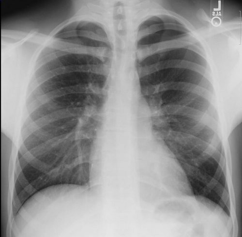
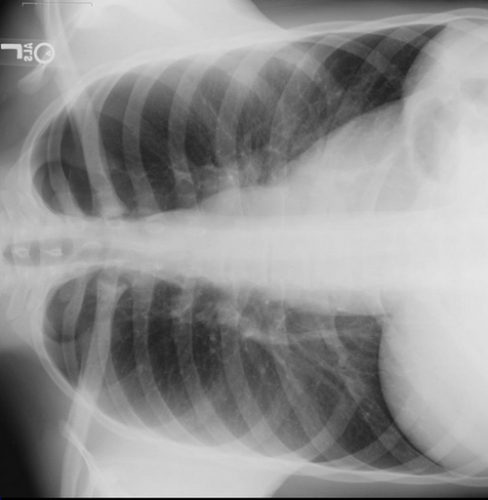
I’ve written previously about how often imaging gets repeated once a trauma patient gets transferred to a trauma center (click here). There are many reasons, including clinical indications, need for advanced imaging (reconstructions), or lack of contrast. But at least 20% have to be repeated because the media is incompatible or not sent with the patient. Sounds like a problem, but is it a significant one?

A recent retrospective analysis of about 2,000 transfers to a Level I center looked at the reasons for repeat imaging and changes in outcome due to it. The paper found several interesting things:

  • Repeat imaging was more likely in more severely injured patients
  • Hospitals that transferred more patients to the trauma center tended to do more scans before transfer
  • Patients who had repeat imaging stayed in the ED longer waiting for definitive disposition
  • Repeat images did not improve outcomes (LOS, DC home, mortality)
  • A rough estimate of $354 more in charges was attributed to repeat imaging

Bottom line: Repeat imaging is wasteful, expensive and increases time in the ED. And don’t forget about the radiation exposure. With all the emphasis on pushing hospitals to use an electronic medical record, there needs to be a similar push to standardize methods for transferring radiographic images between hospitals to address the problem of repeat imaging.

Related posts:

Reference: Repeat imaging in trauma transfers: A retrospective analysis of computed tomography scans repeated upon arrival to a Level I trauma center. J Trauma 72(5):1255-1262, 2012.

Answer: Finding Rib Fractures On Chest Xray

There was a lot of chatter regarding my practical tip yesterday, rotating the chest xray to better visualize rib fractures. Here’s the quiz xray from yesterday: 

And here’s the lateral view:

The fracture is perfectly placed on the most lateral aspect of the left 9th rib. You can download the full size rotated jpg here if you are having a hard time seeing it on the reduced size image above. Piece of cake!

Related post:

Practical Tip: Finding Rib Fractures On Chest Xray

Here’s a neat trick for finding hard to see rib fractures on standard chest x-rays.

First, this is not for use with CT scans. Although chest CT is the “gold standard” for finding every possible rib fracture present, it should never be used for this. Rib fractures are generally diagnosed clinically, and they are managed clinically. There is little difference in the management principles of 1 vs 7 rib fractures. Pain management and pulmonary toilet are the mainstays, and having an exact count doesn’t matter. That’s why we don’t get rib detail x-rays any more. We really don’t care. Would you deny these treatments in someone with focal chest wall pain and tenderness with no fractures seen on imaging studies? No. It’s still a fracture, even if you can’t see it.

So most rib fractures are identified using plain old chest xray. Sometimes they are obvious, as in the image of a flail chest below.


But sometimes, there are only a few and they are hard to distinguish, especially if the are located laterally. Have a look at this image:


There are rib fractures on the left side side on the posterolateral aspects of the 4th and 5th ribs. Unfortunately, these can get lost with all the other ribs, scapula, lung markings, etc.

Here’s the trick. Our eyes follow arches (think McDonald’s) better than all these crazy lines and curves on the standard chest x-ray. So tip the x-ray on its side and make those curves into nice arches, then let your eyes follow them naturally:


Much more obvious! In the old days, we could just manually flip the film to either side. Now you have to use the rotate buttons to properly position the digital image.

Final exam: click here to view a large digital image of a nearly normal chest xray. There is one subtle rib fracture. See if you can pick it out with this trick. You’ll have to save it so you can manipulate it with your own jpg viewer. If you find it, tweet it out to me! Let’s see who gets it first!

Related posts:

How Much Radiation is the Trauma Team Really Exposed To?

Okay, so you’ve seen “other people” wearing perfectly good lead aprons lifting them up to their chin during portable xrays in the trauma bay. Is that really necessary, or is it just an urban legend?

After hitting the medical radiation physics books (really light reading, I must say), I’ve finally got an answer. Let’s say that the xray is taken in the “usual fashion”:

  • Tube is approximately 5 feet above the xray plate
  • Typical chest settings of 85kVp, 2mAs, 3mm Al filtration
  • Xray plate is 35x43cm

The calculated exposure to the patient is 52 microGrays. Most of the radiation goes through the patient onto the plate. A very small amount reflects off their bones and the table itself. This is the scatter we worry about.

So let’s assume that the closest person to the patient is 3 feet away. Remember that radiation intensity diminishes as the square of the distance. So if the distance doubles, the intensity decreases to one fourth. By calculating the intensity of the small amount of scatter at 3 feet from the patient, we come up with a whopping 0.2 microGrays. Since most people are even further away, the dose is much, much less for them.

Let’s put it perspective now. The background radiation we are exposed to every day (from cosmic rays, brick buildings, etc) amounts to about 2400 microGrays per year. So 0.2 microGrays from chest xray scatter is less than the radiation we are exposed to naturally every hour!

The bottom line: unless you need to work out you shoulders and pecs, don’t bother to lift your lead apron every time the portable xray unit beeps. It’s a waste of time and effort!