Tag Archives: xray

Repeat Imaging: What Good Is It?

I’ve written previously about how often imaging gets repeated once a trauma patient gets transferred to a trauma center (click here). There are many reasons, including clinical indications, need for advanced imaging (reconstructions), or lack of contrast. But at least 20% have to be repeated because the media is incompatible or not sent with the patient. Sounds like a problem, but is it a significant one?

A recent retrospective analysis of about 2,000 transfers to a Level I center looked at the reasons for repeat imaging and changes in outcome due to it. The paper found several interesting things:

  • Repeat imaging was more likely in more severely injured patients
  • Hospitals that transferred more patients to the trauma center tended to do more scans before transfer
  • Patients who had repeat imaging stayed in the ED longer waiting for definitive disposition
  • Repeat images did not improve outcomes (LOS, DC home, mortality)
  • A rough estimate of $354 more in charges was attributed to repeat imaging

Bottom line: Repeat imaging is wasteful, expensive and increases time in the ED. And don’t forget about the radiation exposure. With all the emphasis on pushing hospitals to use an electronic medical record, there needs to be a similar push to standardize methods for transferring radiographic images between hospitals to address the problem of repeat imaging.

Related posts:

Reference: Repeat imaging in trauma transfers: A retrospective analysis of computed tomography scans repeated upon arrival to a Level I trauma center. J Trauma 72(5):1255-1262, 2012.

Papers To Change Our Practice 2: Radiation Exposure

The second paper I’ll be discussing at the Penn Trauma reunion tomorrow deals with radiation exposure in trauma. Specifically, I’ll be talking about the amount of radiation the patient is exposed to during their initial evaluation. A lot of work is being published on this topic, but the paper I selected took a different and more accurate approach.

The trauma group at Sunnybrook in Toronto measured surface radiation exposure in a group of 172 major trauma patients. Dosimiters were placed on the neck, chest and groin, and were ideally kept there during the entire hospital stay. A software algorithm was used to calculate organ dose based on the surface measurements. This differs from the more commonly used method of counting studies and calculating dose based on published averages of radiation delivery.

The study was weakened by the number of patients that were excluded or who decided to remove their dosimeter at some point. But a number of interesting facts were revealed:

  • Patients received an average of 5 CT scans and 14 plain xrays during their stay
  • The average total effective dose was 23mSv, about 10 times the normal background exposure for an entire year
  • A surprisingly high dose was delivered to the thyroid, which is more sensitive to radiation exposure
  • A total of 190 extra cancer mortalities would be expected per 100,000 patients, given these exposure numbers
  • Radiation was underestimated using non-dosimeter techniques

Bottom line: We know radiation exposure occurs in our patients, and we know that it’s increasing. It won’t be that long until we start to see the after-effects of these imaging studies, especially in younger patients. What you can’t see does hurt your patients! We need to quickly strike a balance between avoiding missed injuries and irradiating the patient. Specific guidelines to direct ordering of radiographic studies must be developed, and our radiology colleagues need to continue to strive for techniques that adhere to the ALARA (as low as reasonably achievable) philosophy.

Related posts:

Reference: Radiation exposure from diagnostic imaging in severely injured trauma patients. J Trauma 62(1):151-156, 2007.

The Deep Sulcus Sign

Pneumothorax is frequently difficult to diagnose in the resuscitation room. Sometimes it is obvious, with a hypoxic patient and absent breath sounds. But not usually. Most of the time we rely on a chest xray to help make the diagnosis.

Unfortunately, the good old chest xray only shows a pneumothorax about 30-50% of the time. A big part of the problem is that our patients are usually supine to protect their spine. A small pneumothorax make float anteriorly in the supine position, and if it is not big enough to wrap around the lateral edge of the lung, it may remain invisible. So you need to look for gross and subtle signs on the image that will help make the diagnosis. The deep sulcus sign is one of the more subtle signs. 

Simply stated, the deep sulcus sign is a radiolucent (dark) lateral sulcus where the chest wall meets the diaphragm. The amount of lung in this area is less, so a small amount of air will tend to darken the area making it more prominent. Look at patient left in the left photo, and compare to their right side. It is much darker and appears to extend lower than usual. In more extreme cases, the amount of air just above the diaphragm may make it appear inverted (right photo).

Bottom line: If you see a deep sulcus sign on the chest xray image, strongly consider pneumothorax. If the patient begins to have hemodynamic problems, needle the chest and chase with a chest tube. If they remain stable, the patient will still require a chest tube. Chest xray always underestimates the true size of the pneumothorax. Place the usual size chest tube and manage per your usual protocol. And, as always, use your best sterile technique and definitively identify the proper side before placing the tube.

Related posts:

Do You Really Need To Repeat That Xray?

It happens all the time. You get that initial chest and/or pelvic xray in the resuscitation room while evaluating a blunt trauma patient. A few minutes later the tech returns with another armful of xray plates to repeat them. Why? The patient was not centered properly and part of the image is clipped.

Do you really need to go through the process of setting up again, moving the xray unit in, watching people run out of the room (if they are not wearing lead, and see my post below about how much radiation they are really exposed to), and shooting another image? The answer to the question lies in what you are looking for. Let’s address the two most common (and really the only necessary) images needed during early resuscitation of blunt trauma.

First, the chest xray. You are really looking for 3 things:

  • Big air (pneumothorax)
  • Big blood (hemothorax)
  • Big mediastinum (hinting at aortic injury)

Look at the clipped xray above. A portion of the left chest wall is off the image. If there were a large pneumothorax on the left, would you be able to see it? What about a large hemothorax? And the mediastinum is fully included, so no problem there. So in this case, no need to repeat immediately.

The same thing goes for the pelvis. You are looking for gross disruption of the pelvic ring, especially posteriorly because this will cause you to intervene in the ED (order blood, consider wrapping the pelvis). So if parts of the edges or top and bottom are clipped, no big deal.

Bottom line: Don’t let the xray tech disrupt the team again by reflexively repeating images that are not technically perfect. See if you can use what you already have.  And how do you decide if you need to repeat it later, if at all? Consider the mechanism of injury and the physical exam. Then ask yourself if there is anything you could possibly see that was not imaged the first time that would change your management in any way. If not, you don’t need it. But it certainly will irritate the radiologists!

Related posts: 

Pelvic Trauma Radiographs Demystified

Although we are becoming increasingly reliant on CT scans for diagnosis, plain old radiographs still have their place. This is especially true in pelvic imaging after trauma. 

The most common pelvic radiograph obtained is the supine A-P view taken during trauma resuscitation. This image gives a quick and dirty look at the entire pelvis, from iliac crest to ischial tuberosity. The main areas of interest are the pubic symphysis and the SI joints, so if some of the periphery is cut off a repeat is not necessary prior to CT scan. This image helps predict the need for blood and pelvic compression devices.

If fractures are present, the orthopedic surgeons will generally request additional views in addition to the CT scan. The scan gives excellent detail, but the axial image slices are still not as good as a plain old radiograph in many cases.

Inlet and outlet views are used to get a better look at the pelvic ring. The inlet view opens the ring up into a big circle (or oval) and allows identification of fractures of the sacrum or displacement of the SI joints, as well as changes in the pubic symphysis. The outlet view shows any vertical displacements through the sacrum or SI joints well, and gives a better appreciation of some pubic fractures.

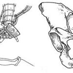
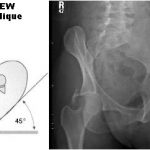
Judet views help demonstrate acetabular fractures by lining up the iliac wing with the xray tube. They can give additional information that the orthopedists use for determining operative or nonoperative management.

Rule of thumb: For major trauma patients, obtain an A-P pelvis radiograph if indicated by mechanism of injury or physical exam. Perform CT scan of the abdomen and pelvis if indicated. If a pelvic ring fracture is identified, obtain inlet and outlet radiographs before calling your orthopedic surgeon. If an acetabular fracture is seen, obtain Judet views before calling.