Tag Archives: what the heck?

What’s With Those John / Jane Doe Names?

Oftentimes, trauma patients arrive, are not very responsive, and are not carrying an ID.  However, our electronic health record systems have a very difficult time with this. To expedite care, most hospitals developed a system of pseudo-names to be used until the patient can be identified.

Originally, these names were often “Doe” names. The classic ones were John Doe and Jane Doe. Obviously, more than two names were needed, so other first names were adopted to provide a sizable pool of pseudo-names.  Other disciplines, such as law enforcement, have used a similar system.

Where did the concept of John/Jane Doe come from? The origin is the British legal system, way back in the 18th century. In those days, landlords would start a “process of ejectment,” known these days as eviction, to rid their properties of landlords or deadbeat tenants. The legal process was a bit complex, so they would file for the process using a fake name to initiate it. They frequently selected John Doe or Richard Roe for their filings.

The reason the Doe and Roe names were used has been lost to history. There is speculation that these names were derived from certain deer species endemic to Britain. But honestly, no one knows for sure now.  Although this process was dropped in Great Britain in the mid-1800s, it persists in the US legal system. Remember the landmark legal case Roe v Wade?

The use of Doe names in electronic health records is rapidly fading as well. The list of name pairs ending with Doe is just not unique enough. There are too many opportunities to mix up similar names, especially if the last one is always the same. This can result in catastrophic errors if test results are misinterpreted, or a blood transfusion with incorrect ABO typing is given to the wrong patient.

Most trauma centers have adopted temporary naming systems consisting of two words or unique names. Some use number and letter sequences combined with another unique word. The real trick in the electronic medical record world is smoothly merging the records utilizing the pseudo-name with the patient’s previous records under their actual name.

The best practice for this varies by electronic record system and hospital. If done too early, the change may disrupt critical processes, such as the massive transfusion protocol. If done too late, it is difficult for trauma professionals to see any records the patient may have under their actual name. Each center must develop its own system for converting from John Doe to the real name.

What The Heck! You Make The Diagnosis – The Answer

In my last post, I detailed the following case:

This male patient was brought to the trauma center after a high-speed car crash. He was unresponsive with GCS 3. A bleeding facial laceration was present, as was vomitus in the airway.

Prehospital providers rapidly intubated the patient and inserted an orogastric tube. They rapidly packaged and transported him to the nearest trauma center.  The facial laceration was stapled for bleeding control. The airway was checked with a CO2 color change indicator and was positive. OG was hooked to suction with return of gastric contents.

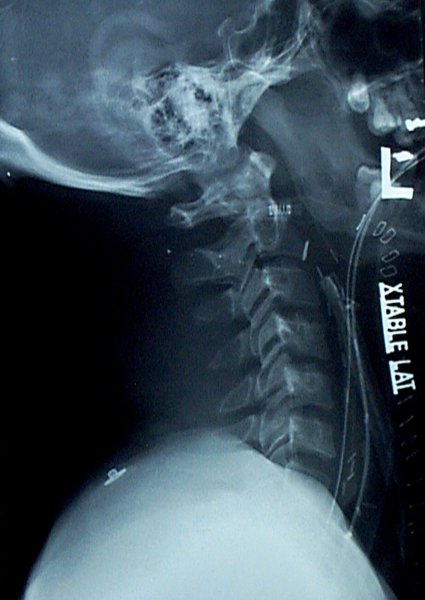
And here was a lateral cervical spine image:

The main problem is that, if you look closely there are three tubes on the image!

Look carefully at the anterior pharynx and trace the radiopaque markers back. There are two nearly overlapping lines. One extends posteriorly, down into the esophagus. This is the orogastric tube. The distal tip of the other (an endotracheal tube which is only inserted to 12 cm at the teeth) stops where it touches another tube. Another endotracheal tube, the proximal end of which is sitting at the epiglottis!

What the heck??! The medics were interviewed, and the patient was initially intubated successfully. The intubator turned his attention to finding a tube securing device, and when he turned back the tube was gone! So he intubated again but met some resistance. This explained the shallow position of the tube.

The patient was oxygenated well and the “outside” ET tube was removed. Then ring forceps were passed under direct vision and the “inside” tube was removed. A well-positioned ET tube was then reinserted. The patient did well afterwards.

Teaching point: When inserting anything that is partially in and partially out of the body (e.g. guidewires, and now ET tubes) always anchor them with your fingers so they don’t just “disappear.” And if you need more hands, ask for assistance!

Source: personal collection. Not treated at Regions Hospital or even in Minnesota.

What The Heck! You Make The Diagnosis

Please help figure out what is wrong here. I’m not going to give you much information, though.

This male patient was brought to the trauma center after a high-speed car crash. He was unresponsive with GCS 3. A bleeding facial laceration was present, as was vomitus in the airway.

Prehospital providers rapidly intubated the patient and inserted an orogastric tube. They rapidly packaged and transported him to the nearest trauma center.  The facial laceration was stapled for bleeding control. The airway was checked with a CO2 color change indicator and was positive. OG was hooked to suction with return of gastric contents.

This case occurred in the old days when lateral cervical spine films were initially used to evaluate the c-spine in the trauma bay. Here is the image:

What is your diagnosis? And what does the team need to do?

Please post your answer in the Disqus comment box below, or email me. You might consider Twitter (or is it X now), but it seems somewhat flaky.

Answers in my next post!

Source: personal collection. Not treated at Regions Hospital or even in Minnesota.


What The Heck?! The Answer!

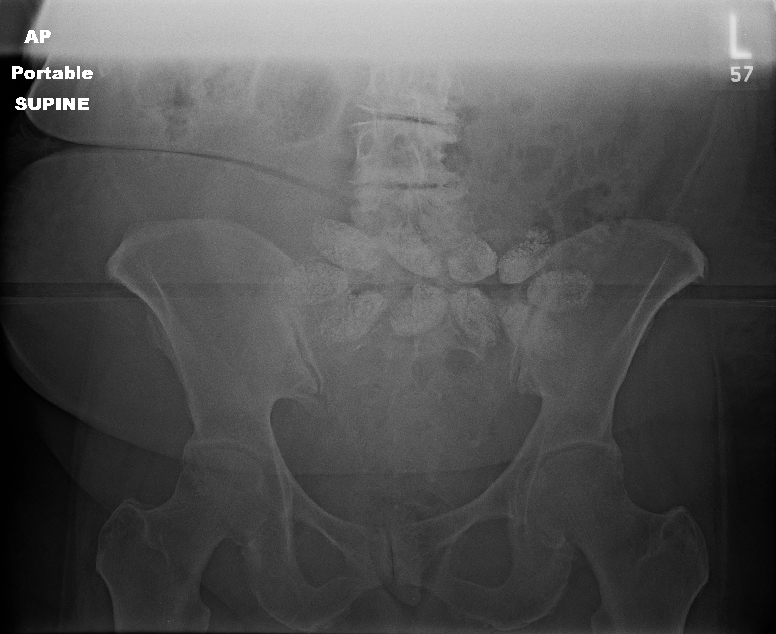
In my last post, I described an elderly pedestrian struck by a car. During the trauma activation, routine chest and pelvic x-rays were obtained. Here was the pelvic image:

Note the odd oval densities across the center of the x-ray. What the heck? What are they?

There are two choices: they are either inside or outside the patient. We had already removed her clothes, so it wasn’t something she was carrying. And if it was inside, we would be able to identify it on the CT scan we had ordered.

But in this case, the x-ray was done early in the secondary survey. Specifically, we did it before we rolled our patient and examined her back.  When we did, here is what we found:

Only it wasn’t in the box. Or on her neck. This one was stuck on her lower back, but not in her clothes. She was suffering from lower back pain, and applied one of these on a daily basis for comfort. We had not rolled the patient prior to the pelvic x-ray.

The pods on these thermal wraps contain a mixture of iron, sodium chloride, sodium thiosulfate, water, charcoal, and sodium polyacrylate that heat up when removed from their package and exposed to oxygen. The iron renders it somewhat radio-opaque, hence their appearance on the x-ray. We did peel it off prior to CT since it would probably create a significant amount of scatter which would degrade the image.

Should we have waited a few more minutes to get the image until we had rolled and examined the back? This is a judgment call. Since our trauma team moves quickly, we are typically ready to head to the scanner in 15 minutes. In order to improve overall CT scan throughput, we have adopted a 5-minute advance notice policy.  To accomplish this, we don’t want to wait until the very end of the resuscitation to get x-rays. That would end up slowing down our process.

You may feel differently about the timing of the images, or you may have a different method of sequencing your CT scanner. Whatever works best for you. But remember, all trauma patients need to be completely undressed and all of their surfaces, nooks, and crannies inspected before they leave the emergency department!

What The Heck?!

Here’s an interesting case from my image archives.

An elderly female pedestrian was struck by a car. She was hemodynamically stable. During the course of her evaluation as a trauma activation, her clothes were completely removed. (She was kept nice and warm with infrared warmers.)

Early in the secondary survey, chest and pelvic x-rays were obtained. Here is the pelvis image:

What is wrong in this picture?? Leave comments below or tweet your guesses. I’ll publish the answer Friday.