Tag Archives: tips

In The Next Trauma MedEd Newsletter: More Potpourri!

The July issue of Trauma MedEd will be sent out to subscribers near the end of the month. It will review some topics that I find very interesting, and I hope you will to.

This issue is being released to subscribers by July 30. If you sign up any time before then, you will receive it, too. Otherwise, you’ll have to wait until it goes out to the general public at the end of next week. Click this link right away to sign up now and/or download back issues.

In this issue, learn about:

  • The effect of ambulance deceleration on ICP in head injury patients
  • An interesting technique for sealing vacuum systems applied around external fixators
  • An analysis of thrombotic events following TXA administration
  • The utility of a second head CT in patients taking DOACs
  • And one or two more depending on space available!

As always, this month’s issue will go to all of my subscribers first. If you are not yet one of them, click this link right away to sign up now and/or download back issues.

Antihypertensive Treatment In Acute TBI

Yes, we know high blood pressure can be bad. Over the long term, it can accelerate atherosclerotic heart disease and pound away at the kidneys and brain. And when it is acutely elevated to critical levels, it can lead to stroke.

But is it always bad in trauma? Trauma hurts like hell, so it’s no wonder than many of our patients (not suffering blood loss of course) are hypertensive.  But how often have you seen this scenario occur:

An elderly patient fell from standing, striking her head. She is brought to your ED by ground EMS. She has a GCS of 8 (E1 V3 M4) with a BP of 200/130 and pulse of 56.  This meets your trauma activation criteria and the team assembles to meet the patient.

As you move her onto the bed, one of your colleagues calls out for some nicardipine to control the pressure. Is this a wise move? Remember the First Law of Trauma:

Any anomaly in your trauma patient is due to trauma, no matter how unlikely it may seem.

What else can cause hypertension and bradycardia in your trauma patient? In this case, certainly a subdural or epidural hematoma.

And why is that happening? Because the intracranial pressure is elevated from the space-occupying lesion. Remember the formula for cerebral perfusion pressure (CPP):

CPP = MAP – ICP

Where MAP = mean arterial pressure and ICP = intracranial pressure.  Normally the MAP is around 90 torr and ICP is about 10 torr. Thus, the normal CPP is approximately 80. The range is 60  to well over 100 torr, and flow autoregulation keeps brain perfusion constant over this range.

But let’s say that we are psychic and know the ICP of our patient to be 60 because of a large subdural hematoma. Her current CPP is 150 – 60 or about 90 torr. What happens if we start her on a nicardipine drip or some other antihypertensive medication? We can certainly normalize the blood pressure to 120/80. But now her CPP drops to 90  – 60 = 30 torr!

Congratulations, you have just shut down circulation to her brain!

Bottom line: Think first before calling for antihypertensive medications in patients who may have increased intracranial pressure. You may be sabotaging the only mechanism protecting their brain while you are calling your neurosurgeon for help. Your top priority is to get them to the CT scanner while permitting that pressure. If it turns out that there is no evidence for pathology that would lead to increased ICP, then turn to the antihypertensive agents to help protect against stroke. 

How To MacGyver A Ventilator From Common Parts

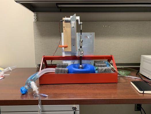
A cardiac anesthesiology fellow, and several engineers from the University of Minnesota and a local device company got together over the past few weeks and cobbled together a ventilator from some spare parts. Here’s a picture to give you an idea of what it looks like:

It uses a metal toolbox tray, an Ambu bag, and some other spare parts lying around one of the medical device labs at the university. Essentially, a servo motor intermittently squeezes the Ambu bag, and there are adjustments for how often (rate) and how deeply (volume) the bag is compressed. There is a pressure limiting device included in the system as well.

This project illustrates how we will need to think outside the (tool)box in the coming weeks, especially as the number of severe Coronavirus cases begins to tax our supply of ventilators. And obviously, this thing will not get FDA approval in your lifetime. But if a choice needs to be made between using something like this in a pinch vs letting someone asphyxiate, the answer is pretty clear.

The group has produced a short YouTube video as well (see below), but it is rather short on details. You get to see some partial views of it as it is being tested on pigs. But so far, the concept is promising.

If you are trying to view this on my Tumblr feed, the video will not show here. Please click here to visit my main blog site to view it.

YouTube player

Thoughts On Traumatic Hematuria: Part 2

Yesterday, I discussed blood in the urine from a urethra. As I mentioned, there is typically not much from that particular injury. Today, I’ll dig into the three causes of real hematuria.

All of these tubes show gross hematuria except the one on the right.

  • Bladder injury. This can occur with either blunt or penetrating injury. The degree of hematuria is variable with stabs or gunshots, but tends to be much darker in blunt injury. This happens because the size of the bladder injury tends to be greater with blunt force. The bladder injury is not necessarily full-thickness with blunt trauma. It may just be some wall contusion and underlying mucosal injury. But frequently, with seat belt injury and/or A-P compression injuries to the pelvis (“open book”), the injury is full thickness.
    • Tip: If less than 50cc of very dark urine flow from the catheter upon insertion, it is likely that your patient has an intraperitoneal bladder rupture!
  • Ureteral injury. This injury is very rare. The most common mechanism is penetrating, but this structure is so small and deep that it seldom gets hit by naything. Patients with multiple lumbar transverse process fractures will occasionally have a small amount of hematuria, probably from a minor contusion. More often than not, the hematuria is microscopic, so we should never know about it.
  • Kidney injury. The most important fact regarding renal injury is that the degree of injury has no correlation with the amount of hematuria. The most devastating injury, a devascularized kidney, frequently has little if any gross hematuria. And conversely, a very minor contusion can produce very red urine.

So what about diagnosis? It’s easy! If you see gross hematuria, insert a foley catheter (if not already done) and order a CT of the abdomen/pelvis with contrast, as well as a CT cystogram. The latter must not be done using passive filling of the bladder with a clamped catheter. Contrast must be infused into the bladder under pressure to ensure a bladder injury can be identified.

CT scan is an excellent tool for defining injuries to kidney, ureter, and bladder, and will identify extravasation into specific places and allow grading. Specific management will be the topic of future posts.

Thoughts On Traumatic Hematuria: Part 1

I’ve seen a number of patients recently with bloody urine, and that is prompting me to provide some (written) clarity to others who need to manage this clinical problem. I’ll try to keep it organized!

There are two kinds of hematuria in trauma: blood that you can see with the naked eye, and…

Okay, so there’s only one. Trauma professionals do not care about microscopic hematuria. It does not change clinical management. Sure, your patient might have a renal contusion, but you won’t do anything about that. Or, he/she might have an infarcting kidney. And you can’t do anything about that. If you order a urinalysis, you might see a few RBCs. Don’t let this lead you down the path of looking for a source. You’ll end up ordering lots of tests and additional imaging, and generally will have nothing to show for it at the end. It’s not your job to spend good money on the very rare chance of finding something clinically significant.

Both of these specimens have blood in them. You can’t see it on the left, so don’t go looking for it with a microscope.

There are four sources of blood in the urine.

1. The first source does not generally cause hematuria, but can occasionally cause a few visible wisps of blood. That source is a urethral injury. The textbook teaching, and it’s good advice, is to look at the urethral meatus in your trauma patient, especially if you are contemplating insertion of a urinary catheter. If you see a few drops of blood, pause to consider. Sometimes, the blood is no longer visible, but might be present as a few well-placed drops on the patient’s underwear. So have a look at that, too, especially in patients with high risk injuries such as A-P compression pelvic fractures (think, lots of ramus fractures or pubic diastasis).

If you didn’t notice it and inserted the catheter anyway, you might see a few wisps of blood in the tubing as you place it. More often than not, this is just run of the mill irritation of the mucosa by the catheter, but always keep the possibility of an injury in mind.

Tomorrow, I’ll discuss the remaining three sources, and what to do about them.

Related posts: