Tag Archives: spleen

Angioembolization For Splenic Injury

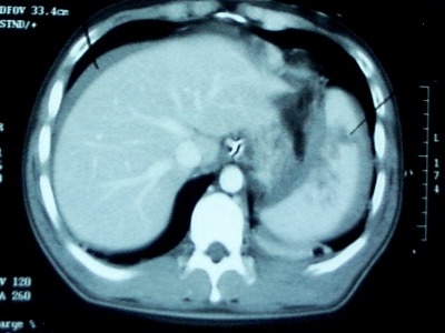
Initial nonoperative management of splenic injury is standard in hemodynamically stable patients. Over the past decade, the success rates have climbed by adding angioembolization to the algorithm, according to several published series. However, the objective benefit and specific indications have not been worked out.

A paper published this month by the University of Florida, Jacksonville used the NTRACS registry to try to clarify these issues. They identified 1039 patients undergoing nonoperative management (NOM) over a nearly 10 year period. Patients who died shortly after arrival, those who went directly to OR for hemodynamic reasons, and children were excluded, leaving 539 patients. Only about 1/6 of the patients underwent embolization. 

The overall failure rate was about 4%, a little higher in the non-angio patients, a little lower with angio. Incidentally, the angio group had significantly higher injury severity (26 vs 20). Analysis of the lower grade spleen injury group showed no improvement in success rate by adding angio. However, the high grade groups (grades IV-V) did benefit by adding this procedure. Similarly, success improved when performing angio in patients with contrast blush or evidence of slow, ongoing bleeding. If NOM did fail, it usually occurred on day 2.

Bottom line: Although we’ve been adding angio to non-operative management of spleen and liver injury for a decade, here’s the first paper that has been able to define the real indications for doing it. First, all unstable patients go to the OR (don’t even consider nonop management). In the remaining patients, if the CT shows a grade IV or V injury, or a contrast blush, angio is recommended. If neither of these is noted, but the hemoglobin continues to decline “too quickly” (surgeon judgement), then a trip to angio is also warranted. Applying these principles can increase your success rate to about 96%.

Related post:

Reference: Selective angiographic embolization of blunt splenic traumatic injuries in adults decreases failure rate of nonoperative management. J Trauma 72(5):1127-1134, 2012.

Delayed Splenic Rupture: Part 2

Yesterday I wrote about the history of “delayed splenic rupture.” Today I’ll discuss how to deal with it.

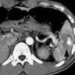
If possible, try to avoid ever having to mess around with this clinical problem. If you order an abdominal CT after blunt trauma and see a splenic contrast blush of either type (pseudoaneurysm or extravasation, see left photo), then deal with it before the patient even knows he has a problem. A trip to interventional radiology will usually solve the problem. And if embolized, these patients almost never come back with a bleeding problem.

As I’ve said many times before, if the patient is hemodynamically compromised, then an OR visit is required. The usual solution is splenectomy. Some recommend repairing the spleen, but this is technically more difficult than it sounds, and it is difficult for the surgeon to sleep soundly after performing one of these.

Lets say you inherited one of these from someone else, or ignored the warning signs on the initial CT. The usual time frame for presentation to the ED with acute bleeding is 7 to 10 days after the initial injury. If they are not stable, physical exam or FAST will quickly direct you to the OR, once again for splenectomy. Some patients will stabilize with fluids and can safely be sent to CT scan.

Once the CT confirms what the problem is, a trip to interventional radiology is in order if the patient remains stable. Here is the key: the radiologist must embolize something! If they find a bleeding vessel, then they can selectively embolize it. If they don’t, then the main splenic artery should be embolized. This will decrease the arterial pressure head, but won’t eliminate it. It will decrease the likelihood of additional bleeding as much as possible.

At this point, the patient should be admitted to the trauma service and monitored using your solid organ injury protocol. If they have any hemodynamic issues, it’s time to remove the spleen. Remember, this is the third time they’ve had a problem, and like in baseball, their spleen is out! Attempted splenorrhaphy at this point is pointless and may lead to yet another operation.

Related posts:

Delayed Splenic Rupture: Part 1

This post was prompted by a paper that somehow got into the Journal of Trauma this month on nonoperative management of delayed splenic rupture after trauma. It’s a bad retrospective review of 15 patients which I’ll say more about tomorrow. There’s very little good literature on this topic, so I wanted to share some personal observations.

Back in the days before CT scan (and unfortunately, I remember them), the diagnosis of abdominal injury was much more difficult. It was primarily qualitative, meaning that we somehow figured out that they either had it or they didn’t. We could not very easily figure out what specific injuries a given patient had. However, management was simple: we went to the operating room, found out and fixed it.

Sometimes, though, we would encounter a patient who had been involved in some type of blunt trauma a week or two earlier who presented to the ED with left-sided abdominal pain, shock and anemia. The diagnosis was “delayed splenic rupture” and they were taken to OR for a splenectomy.

When CT scan came along, we found out that these were actually “delayed recognition of splenic injury.” We still took them to the OR for splenectomy, but with experience this slowly gave way to splenic repair, and then to nonoperative management. 

There is still one subset of these injuries that is problematic: spleen injury with a contrast blush. It turns out that there are really two types of blush: contrast seen within a pseudoaneurysm within the splenic pulp, and extravasation. And furthermore, the pseudoaneurysm is the culprit in most “delayed splenic ruptures.”

Tomorrow, I’ll write about how to recognize this potential problem, what to do about it acutely, and what to do if it was missed and the patient presents to your ED ten days later in shock.

Related post:

Reference: Nonsurgical management of delayed splenic rupture after blunt trauma. J Trauma 72(4):1019-1023, 2012.

When to Give Spleen Vaccines After Splenectomy for Trauma

I’ve written previously on the (f)utility of giving vaccines after splenectomy for trauma (click here to read). However, it is more or less a medicolegal standard, so pretty much everyone gives them. The big question is, when? 

Some centers give them immediately postop, some before hospital discharge, and some during their postop visit. Who is right? The argument is that major surgery produces some degree of immunocompromise. So if the vaccines are given too early, perhaps the anitbodies will not be processed as effectively, and the response to an actual bacterial challenge might not be as good.

One prospective study randomized patients to receive their pneumococcal vaccine either 1, 7, or 14 days after surgery. IgG levels were measured before vaccination and again after 4 weeks. This study found that antibody concentrations were the same in all groups. However, functional activity of the antibodies was low in the 1 and 7 day groups, and nearly normal in the 14 day group.

Following this, a rat study looked at vaccination timing followed by exposure to pneumococcus. These animals were splenectomized, then given a real or sham vaccination at 1, 7, or 42 days. They then had pneumococcus injected into their peritoneal cavity. About 70% of all rats with sham vaccination died. Only 1.5% of the vaccinated rats died, and there were no differences based on vaccination timing.

Bottom line: Neither antibody titer studies nor rat studies easily translate into recommendations for treating overwhelming post-splenectomy sepsis (OPSS) in humans. And such a study can never be done because of the rarity of this condition (less than 70 cases since the beginning of time). It really boils down to your specific population, balancing your assurance that your patient will get it against the possibility that their immune system may not react to it as much as it could. 

At our center, we give the vaccines as soon as possible postoperatively. This ensures that it is given, and erases any doubt of what might happen if the patient does not show up for their postop check.

References:

  • Immune responses of splenectomized trauma patietns to the 23-valent pneumococcal polysaccharide vaccine at 1 versus 7 versus 14 days after splenectomy. J Trauma 44(5):760-766, 1998.
  • Timing of vaccination does not affect antibody response or survival after pneumococcal challenge in splenectomized rats. J Trauma 45(4):682-697, 1998.

Related posts:

    EAST Guidelines Update: Spleen Injury

    The Eastern Association for the Surgery of Trauma is in the process of updating their trauma practice guidelines for spleen injury. The first set of guidelines was introduced in 2003, and several advances in management have occurred since. here is a summary of the current status of the guidelines:

    Level I recommendations (best quality data): 

    • none

    Level II recommendations (good data):

    • Initial management of hemodynamically stable patients should be nonoperative
    • Unstable patients should undergo immediate operation or angiographic embolization (my interpretation: unstable patients belong in the OR, not the angio suite!)
    • Patients with peritonitis should go to the operating room
    • Age, grade of injury, amount of hemoperitoneum and age are not contraindications to nonoperative management. Only hemodynamic stability matters.
    • CT of the abdomen with IV contrast is the most reliable method to assess severity of spleen injury (my interpretation: in the hemodynamically stable patient)
    • Angiography with embolization should be considered if a contrast blush is seen on CT, AAST grade > 3, moderate hemoperitoneum is present, or there is evidence of ongoing bleeding
    • Nonoperative management should only be considered if continuous monitoring and serial exams can be carried out at your hospital, and if an operating room is immediately available if needed

    Level III recommendations (weak data):

    • Clinical status should dictate need and frequency of followup imaging (my interpretation: only do it if the patient condition changes for the worse)
    • Contrast blush is not an absolute indication for operation or angio-embolization. Age, grade of injury and presence of hypotension need to be considered. (My interpretation: don’t operate or do angio on kids without a really good reason)
    • Angio is an adjunct to nonop management in patients who are at high risk for delayed bleeding or to look for vascular injuries (pseudoaneurysms) that may lead to rupture or delayed hemorrhage

    Reference: Trauma Practice Guideline Update, 24th Annual Scientic Assembly, Eastern Association for the Surgery of Trauma, January 2011.