Tag Archives: spleen

Spleen Embolization After Trauma

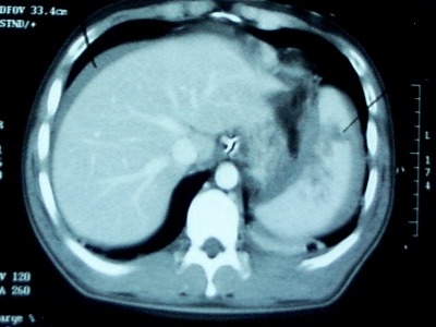
Angioembolization of the spleen (AES) is part of our armementarium in the management of spleen and liver trauma. However, there are no good guidelines to help us decide exactly which patients would benefit from it. An abstract to be presented at the EAST meeting in January 2013 gives us a little more information on the actual benefits of this procedure.

The authors did a retrospective review of the management of blunt splenic injury at four busy Level I trauma centers. They looked at 1275 injured patients over a 3 year period. Here are the interesting tidbits from the study:

  • There was considerable variation in the use of AES at the 4 centers, ranging from 1% of patients to 14%. This should be no surprise because there is no real guidance available yet.
  • There was also a large degree of variation between the number of initial splenectomy performed at these centers
  • Centers that used AES more frequently had lower initial splenectomy rates
  • Patients at centers with high AES rates were 3 times more likely to leave with their spleen intact

Bottom line: This abstract correlates with my own personal experience: judicious use of angioembolization saves spleens. The real question is about which patients are best served by it. Our protocol is to strongly consider it in all high grade spleen injuries (Grade 4 and 5), and to always do it if a blush or extravasation is present. Our success rate for nonoperative management currently stands at about 94%.

Related posts:

Reference: Variation in splenic artery embolization a spleen salvage: a multicenter analysis. EAST Annual Scientific Assembly, Paper #1, to be presented January 2013.

When to Give Spleen Vaccines After Splenectomy for Trauma

I’ve written previously on the (f)utility of giving vaccines after splenectomy for trauma (click here to read). However, it is more or less a medicolegal standard, so pretty much everyone gives them. The big question is, when? 

Some centers give them immediately postop, some before hospital discharge, and some during their postop visit. Who is right? The argument is that major surgery produces some degree of immunocompromise. So if the vaccines are given too early, perhaps the anitbodies will not be processed as effectively, and the response to an actual bacterial challenge might not be as good.

One prospective study randomized patients to receive their pneumococcal vaccine either 1, 7, or 14 days after surgery. IgG levels were measured before vaccination and again after 4 weeks. This study found that antibody concentrations were the same in all groups. However, functional activity of the antibodies was low in the 1 and 7 day groups, and nearly normal in the 14 day group.

Following this, a rat study looked at vaccination timing followed by exposure to pneumococcus. These animals were splenectomized, then given a real or sham vaccination at 1, 7, or 42 days. They then had pneumococcus injected into their peritoneal cavity. About 70% of all rats with sham vaccination died. Only 1.5% of the vaccinated rats died, and there were no differences based on vaccination timing.

Bottom line: Neither antibody titer studies nor rat studies easily translate into recommendations for treating overwhelming post-splenectomy sepsis (OPSS) in humans. And such a study can never be done because of the rarity of this condition (less than 70 cases since the beginning of time). It really boils down to your specific population, balancing your assurance that your patient will get it against the possibility that their immune system may not react to it as much as it could. 

At our center, we give the vaccines as soon as possible postoperatively. This ensures that it is given, and erases any doubt of what might happen if the patient does not show up for their postop check.

References:

  • Immune responses of splenectomized trauma patietns to the 23-valent pneumococcal polysaccharide vaccine at 1 versus 7 versus 14 days after splenectomy. J Trauma 44(5):760-766, 1998.
  • Timing of vaccination does not affect antibody response or survival after pneumococcal challenge in splenectomized rats. J Trauma 45(4):682-697, 1998.

Related posts:

DVT Prophylaxis After Solid Organ Injury

Nonoperative management of solid organ injury is the norm, and has reduced the operative rate significantly. At the same time, the recognition that development of deep venous thrombosis (DVT) in trauma patients is commonplace creates uncertainty? Is it safe to give chemical prophylaxis with low molecular weight heparin (LMWH)? How soon after injury?

The trauma group at USC+LAC published the findings of a retrospective review of 312 patients undergoing nonoperative management for their liver, spleen or kidney injuries. They looked at chemical prophylaxis administration and its relationship to failure of nonop management of solid organ injury.

As expected, as the grade of the solid organ injury increased, so did the failure rate of nonoperative management. Administration of low molecular weight heparin, such as enoxaparin, did not increase failure rate in this study. All but one failure occurred in patients who had not yet received the injections. Likewise, two DVT and two pulmonary embolisms occurred, but only in patients who had not yet received prophylaxis. 

Bottom line: This small study offers some assurance that early prophylaxis is okay, and a few prospective studies do exist. UCSF / San Francisco General is comfortable beginning chemical prophylaxis 36 hours postop, regardless of solid organ injury. Look for more guidance on this issue in the near future. Until then, consider starting LMWH prophylaxis early to avoid complications from DVT or PE.

Reference: Thromboembolic prophylaxis with low-molecular-weight heparin in patients with blunt solid abdominal organ injuries undergoing nonoperative management: current practice and outcomes. J Trauma 70(1): 141-147, 2011.

Undergrading Spleen Injury

We love our CT scans! They’re so high tech, with such detailed images popping up on the monitor so quickly. To take advantage of the detail, we’ve come up with fancy grading systems that can be used to direct care. But are they all they’re cracked up to be?

CT grading of spleen injury is a prime example. We’ve got a nice, detailed system that looks at laceration depth, subcapsular hematoma size and vascular injury. We can use it to predict the likelihood of needing an operation and where we should admit someone in the hospital (ICU vs ward). And when we see the injury on the screen, we believe that we can accurately apply the scoring system to these beautiful images.

But unfortunately, it’s not that simple.Scanning obtains multiple images in an axial plane and lays them out for us to look at. However, the spleen (and most other organs) and not shaped like a cube. It is curved, with complex nooks and crannies that can look like cracks. Moderate to large hematoma around the spleen can obscure lacerations. And the hilum is even more complicated and variable in shape.

Because of this, CT scans of the abdomen tend to underestimate the true extent of injury, especially in the higher grades. Grade I and II injuries are usually accurate, but in Grades III-V, the scan tends to undergrade by 1 (30% of cases) or 2 grades (45% of cases) when re-graded at surgery.

Bottom line: Grade I and II injuries are generally managed in a lower intensity setting and almost never require operation. But beware of the higher grades! It is very likely that it’s higher than you think. This means that if your patient slowly becomes tachycardic or their blood pressure softens, believe the clinical evidence. Don’t rely on a CT scan that was done hours ago that may be hiding a more severe injury than you think! (This applies to liver injuries as well)

Related posts:

Reference: Correlation of operative and pathological injury grade with computed tomographic grade in the failed nonoperative management of blunt splenic trauma. Euro J Trauma Emerg Surg – Online First 2 Mar 2012.

Angioembolization For Splenic Injury

Initial nonoperative management of splenic injury is standard in hemodynamically stable patients. Over the past decade, the success rates have climbed by adding angioembolization to the algorithm, according to several published series. However, the objective benefit and specific indications have not been worked out.

A paper published this month by the University of Florida, Jacksonville used the NTRACS registry to try to clarify these issues. They identified 1039 patients undergoing nonoperative management (NOM) over a nearly 10 year period. Patients who died shortly after arrival, those who went directly to OR for hemodynamic reasons, and children were excluded, leaving 539 patients. Only about 1/6 of the patients underwent embolization. 

The overall failure rate was about 4%, a little higher in the non-angio patients, a little lower with angio. Incidentally, the angio group had significantly higher injury severity (26 vs 20). Analysis of the lower grade spleen injury group showed no improvement in success rate by adding angio. However, the high grade groups (grades IV-V) did benefit by adding this procedure. Similarly, success improved when performing angio in patients with contrast blush or evidence of slow, ongoing bleeding. If NOM did fail, it usually occurred on day 2.

Bottom line: Although we’ve been adding angio to non-operative management of spleen and liver injury for a decade, here’s the first paper that has been able to define the real indications for doing it. First, all unstable patients go to the OR (don’t even consider nonop management). In the remaining patients, if the CT shows a grade IV or V injury, or a contrast blush, angio is recommended. If neither of these is noted, but the hemoglobin continues to decline “too quickly” (surgeon judgement), then a trip to angio is also warranted. Applying these principles can increase your success rate to about 96%.

Related post:

Reference: Selective angiographic embolization of blunt splenic traumatic injuries in adults decreases failure rate of nonoperative management. J Trauma 72(5):1127-1134, 2012.