Tag Archives: spleen

Vaccines After Splenic Trauma

The current standard of care is to vaccinate patients after splenectomy to prevent overwhelming post-splenectomy sepsis (OPSS). The real questions are, is this reasonable and is it needed after splenorrhaphy or angioembolization, too?

The spleen was recognized as contributing to infection resistance in the early 1900s. A study on post-splenectomy sepsis that has been widely quoted was published in 1952. Unfortunately, the children involved all had hematologic disorders, so it is difficult to determine if their sepsis deaths were due to splenectomy or their underlying disease.

Reports of sepsis and death continued to accumulate in the latter half of the last century, but there was a tremendous amount of overlap in patient cases. Richardson reviewed the world literature to date and found that, as of about 2003, there were roughly 70 total cases worldwide since the beginning of time, with a death rate of about 30%. Basically, there are more published papers and reports on death from OPSS than there are actual cases!

This flawed data directed a push toward splenorrhaphy and then to nonoperative management of splenic injury. Guidelines have been developed and revised that suggest that the following vaccines should be given to patients with splenectomy:

  • Pneumovax 23 – .5cc SQ, booster every 6 years
  • Haemophilus B conjugate – .5cc IM, no booster
  • Meningococcal vaccine (polysaccharide or diphtheria conjugate) – .5cc (route depends on vaccine), booster status unclear

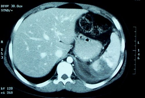
There is no good data at all on vaccine administration after angioembolization. Animal studies suggest that at least 50% of the spleen must be perfused by the splenic artery in order to maintain immune competence. Patients who have CT or angiographic evidence that a significant portion of the spleen is not perfused should probably undergo vaccination.

Given the rarity of OPSS and the even lower probability of dying from it, a definitive study regarding the usefulness of spleen vaccine administration will never be done. So we are stuck with giving them in spleen-injured or spleen-free patients even though the usefulness can never be proven.

Reference: J David Richardson. Managing Liver and Spleen Injuries. J Am Col Surg 200(5):648-669, 2005.

Splenic Vascular Blush

Contrast blush is always a concern when seen on CT of the abdomen for trauma. It can represent one of two things, and both are bad:

  • Active extravasation of contrast
  • Splenic pseudoaneurysm

These two clinical issues can be distinguished by looking at the location of the contrast and its persistence. A pseudoaneurysm is located within the parenchyma, and the contrast will wash away, so it will not be visible on delayed images. Contrast that extends beyond the parenchyma or persists in delayed views represents active bleeding. In either case, the failure rate of nonoperative management exceeds 80% in adults without additional measures being taken.

Clinically, these patients usually act as if they are losing volume and require additional crystalloid and/or blood transfusion. The natural history in adults is for bleeding to continue or for the pseudoaneurysm to rupture, resulting in a quick trip to the operating room.

If vital signs can be maintained with fluids and blood, a trip to interventional radiology may solve the problem. Selective or nonselective embolization can be carried out and patients with only a few bleeding points can be spared operation. However, if multiple bleeding areas are seen, it is probably better to head to the OR for splenorrhaphy or splenectomy.

The image below shows likely areas of extravasation. They are a bit large to be pseudoaneurysms.

Spleen Blush-CT

Children are different than adults. Extravasation from spleen injuries in prepubescent children frequently stops on its own. Angiography should only be used if the child is failing nonoperative management.

Grading Spleen Injuries Simplified

Spleen injury grading is not as complicated as people think! The grading system ranges from Grade I (very minor) to Grade V (shattered, devascularized). 

There is one nuance that people frequently don’t appreciate: multiple injuries can increase the grade. Technically, multiple injuries advance the maximum grade by one point, up to a maximum of Grade 3. So Grade 1 + Grade 1 = Grade 2, but Grades 2+2 = 3! Weird arithmetic!

The vast majority of injuries are Grades 1 to 3, and they are actually the easiest to grade. I use this simple rule: 1 and 3, 10 and 50.

The first set of numbers indicates the depth of a laceration in centimeters.

  • Grade 1 – < 1 cm laceration depth
  • Grade 2 – 1-3 cm laceration depth
  • Grade 3 – >3 cm laceration depth

The second set of numbers refers to size of a subcapsular hematoma in percent of the total surface area of the spleen. Hint: most of these low grades are determined by laceration depth. Very few actually have sizable subcapsular hematomas. So memorize the 1-3 rule first!

  • Grade 1 – <10% subcapsular hematoma
  • Grade 2 – 10-50% subcapsular hematoma
  • Grade 3 – >50% subcapsular hematoma

Grades 4 and 5 use other criteria, but in general if it looks completely pulped it’s a 5, and if it’s a little less pulped, it’s a 4.

  • Grade 4 – hilar injury with >25% devascularization OR contrast blush (active bleeding)
  • Grade 5 – shattered spleen, or nearly complete devascularization

That’s it! Tomorrow I’ll talk about the real significance of the contrast blush.

Spleen Week

This week I’ll be covering spleen injuries. The answer to the question “What is wrong with this spleen” is: 1. There is a spleen laceration (grade cannot be determined from this one slice) and 2. There is a contrast blush.

Today I’ll cover grading and tomorrow I’ll talk about the significance of blushes.