Tag Archives: radiation

How Much Radiation is the Trauma Team Really Exposed To?

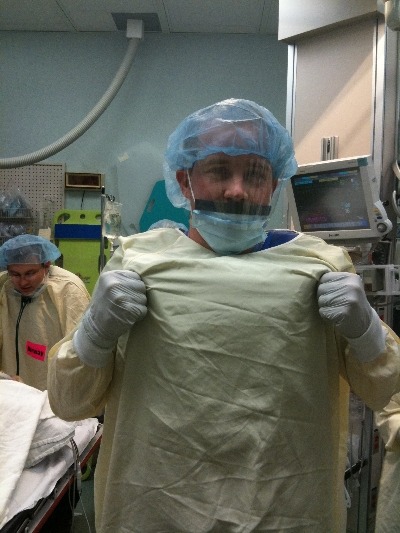
Previously, I posted about “other people” wearing perfectly good lead aprons lifting them up to their chin during portable xrays in the trauma bay. Is that really necessary, or is it just an urban legend?

Lead apron fly

After hitting the medical radiation physics books (really light reading, I must say), I’ve finally got an answer. Let’s say that the xray is taken in the “usual fashion”:

  • Portable technique in your trauma bay
  • Tube is approximately 5 feet above the xray plate
  • Typical chest settings of 85kVp, 2mAs, 3mm Al filtration
  • Xray plate is 35x43cm

The calculated exposure to the patient is 52 microGrays. Most of the radiation goes through the patient onto the plate. A very small amount reflects off their bones and the table itself. This is the scatter we worry about.

So let’s assume that the closest person to the patient is 3 feet away (1 meter). Remember that radiation intensity diminishes as the square of the distance. So if the distance doubles, the intensity decreases to one fourth. By calculating the intensity of the small amount of scatter at 3 feet from the patient, we come up with a whopping 0.2 microGrays. Since most people are even further away, the dose is much, much less for them.

Let’s put it perspective now. The background radiation we are exposed to every day (from cosmic rays, brick buildings, etc) amounts to about 2400 microGrays per year. So 0.2 microGrays from chest xray scatter is less than the radiation we are exposed to naturally in about 44 minutes!

The bottom line: unless you need to work out you shoulders and pecs, don’t bother to lift your lead apron every time the portable xray unit beeps. It’s a waste of time and effort, unless you are dealing with xray imaging on a very regular basis! And that 52 microGrays the patient absorbed? That’s 8 days worth of background radiation.

The Lead Gown Pull-Up: Part 2

Okay, so you’ve seen “other people” wearing perfectly good lead aprons lifting them up to their chin during portable x-rays in the trauma bay. Is that really necessary, or is it just an urban legend?

After hitting the medical radiation physics books (really light reading, I must say), I’ve finally got an answer. Let’s say that the xray is taken in the “usual fashion”:

  • Tube is approximately 5 feet above the xray plate
  • Typical chest settings of 85kVp, 2mAs, 3mm Al filtration
  • Xray plate is 35x43cm

The calculated exposure to the patient is 52 microGrays. Most of the radiation goes through the patient onto the plate. A very small amount reflects off their bones and the table itself. This is the scatter we worry about.

So let’s assume that the closest person to the patient is 3 feet away. Remember that radiation intensity diminishes as the square of the distance. So if the distance doubles, the intensity decreases to one fourth. By calculating the intensity of the small amount of scatter at 3 feet from the patient, we come up with a whopping 0.2 microGrays. Since most people are even further away, the dose is much, much less for them.

Let’s put it perspective now. The background radiation we are exposed to every day (from cosmic rays, brick buildings, etc) amounts to about 2400 microGrays per year. So 0.2 microGrays from chest x-ray scatter is less than the radiation we are exposed to naturally every hour!

The bottom line: unless you need to work out you shoulders and pecs, you probably don’t bother to lift your lead apron every time the portable x-ray unit beeps. It’s a waste of time and effort! Just stand back and enjoy!

Trauma Mortality vs Cancer Mortality from CT Scans for Trauma

Trauma professionals worry about radiation exposure in our patients. A lot. There are a growing number of papers dealing with this topic in the journals every month. The risk of dying from cancer due to CT scanning is negligible compared to the risk from acute injuries in severely injured patients. However, it gets a bit fuzzier when you are looking at risk vs benefit in patients with less severe injuries. Is it possible to quantify this risk to help guide our use of CT scanning in trauma?

A nice paper from the Mayo clinic looked at their scan practices in 642 adult patients (age > 14) over a one year period. They developed dose estimates using a detailed algorithm, and combined them with data from the Biological Effects of Ionizing Radiation VII data. The risk level for injury was estimated using their trauma team activation criteria. High risk patients met their highest level activation criteria, and intermediate risk patients met their intermediate level activation criteria.

Key points in this article were:

  • Average radiation dose was fairly consistent across all age groups (~25mSv)
  • High ISS patients had a significantly higher dose
  • Cumulative risk of cancer death from CT radiation averaged 0.1%
  • This risk decreased with age. It was highest in young patients (< 20 yrs) at 0.2%, and decreased to 0.05% in the elderly (> 60 yrs)

Bottom line: Appropriate CT scan use in trauma evaluation is challenging. It’s use is widespread, and although it changes management it has not decreased trauma mortality. This paper shows that the risk of death from trauma in the elderly outweighs the risk of death from CT scan radiation. However, this gap narrows in younger patients with less serious injuries because of their very low mortality rates. Therefore, we need to focus our efforts to reduce radiation exposure on our young patients with minor injuries.

Related posts:

References:

  • Comparison of trauma mortality and estimated cancer mortality from computed tomography during initial evaluation of intermediate-risk trauma patients. J Trauma 70(6):1362-1365, 2011.
  • Health risks from low levels of ionizing Radiation: BEIR VII, Phase 2. Washington DC: The National Academies Press, 2006.