Tag Archives: radiation

The Lead Gown Pull-Up: Part 3!

Okay, I’ve written about the lead gown pull-up several times.  Here’s how it goes:

I wrote in some detail about when this is necessary for thyroid and thymus protection and how much radiation exposure the trauma team actually gets.

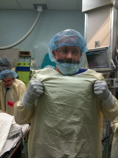
But recently I’ve noticed some members of my own trauma team failing to wear the lead aprons, AND leaving the room when x-rays are taken!

Here’s the thing. Yes, it is important to shield yourself when working in proximity to the x-ray machine when in use. But no, leaving the room is not an acceptable way of accomplishing this! The patient is relatively less attended, and by definition less gets done while several of the team members are outside the room waiting for x-the ray tech to shoot.

Here’s my solution: I make a special announcement as part of the team pre-briefing (before patient arrival) that the lead gown is part of their personal protective equipment (PPE). It is also expected that everybody wears appropriate shielding. We already have a rule that every member of the trauma team MUST wear PPEs or they can’t enter the resuscitation room. And I follow it up by announcing my new rule: if anyone leaves the room because they don’t have proper PPEs, they will not be allowed back in the room. 

Works like a charm!

Related post:

The Lead Gown Pull-Up: Part 2

Okay, so you’ve seen “other people” wearing perfectly good lead aprons lifting them up to their chin during portable x-rays in the trauma bay. Is that really necessary, or is it just an urban legend?

After hitting the medical radiation physics books (really light reading, I must say), I’ve finally got an answer. Let’s say that the xray is taken in the “usual fashion”:

  • Tube is approximately 5 feet above the xray plate
  • Typical chest settings of 85kVp, 2mAs, 3mm Al filtration
  • Xray plate is 35x43cm

The calculated exposure to the patient is 52 microGrays. Most of the radiation goes through the patient onto the plate. A very small amount reflects off their bones and the table itself. This is the scatter we worry about.

So let’s assume that the closest person to the patient is 3 feet away. Remember that radiation intensity diminishes as the square of the distance. So if the distance doubles, the intensity decreases to one fourth. By calculating the intensity of the small amount of scatter at 3 feet from the patient, we come up with a whopping 0.2 microGrays. Since most people are even further away, the dose is much, much less for them.

Let’s put it perspective now. The background radiation we are exposed to every day (from cosmic rays, brick buildings, etc) amounts to about 2400 microGrays per year. So 0.2 microGrays from chest x-ray scatter is less than the radiation we are exposed to naturally every hour!

The bottom line: unless you need to work out you shoulders and pecs, you probably don’t bother to lift your lead apron every time the portable x-ray unit beeps. It’s a waste of time and effort! Just stand back and enjoy!

The Lead Gown Pull-Up?

Trauma Team members typically wear a lead gown under their personal protective equipment so they don’t have to run out of the room when x-rays are taken. How often do you see people do this?

Is it really necessary? Or is it just a way to exercise your pecs and biceps? Tomorrow I’ll talk about how much radiation team members are really exposed to so you can decide if this is really necessary.

How Much Radiation Exposure In Imaging Studies?

Everyone knows that CT scans deliver more radiation than conventional x-ray. But how much does each test really deliver? And how significant is that?

Let me try to put it all into perspective. First, how much radiation are we exposed to just living outside the hospital? Background radiation is everywhere. It consists of radioactive gases (argon) in the air we breathe, radiation from the rocks and other things around us, and cosmic rays blasting through us from space.

In the United States, the average background radiation each of us is exposed to is about 3.1 milliSieverts (mSv). I’ve compiled a table to show the approximate dose delivered by some of the common radiographic studies ordered by trauma professionals. And to keep it real, I’ve calculated how much extra background radiation we would have to absorb, in units of time, to have an equivalent exposure.

Read and enjoy! Remember, doses may vary by scanner, settings, and dose reduction measures used.

Test Dose (mSv) Equivalent background
radiation
Chest x-ray 0.1 10 days
Pelvis x-ray 0.1 10 days
CT head 2 8 months
CT cervical spine 3 1 year
Plain c-spine 0.2 3 weeks
CT chest 7 2 years
CT abdomen/pelvis 10 3 years
CT T&L spine 7 2 years
Plain T&L spine 3 1 year
Millimeter wave
scanner (that hands
in the air TSA thing at
the airport)
0.0001 15 minutes
Scatter from a chest
x-ray in trauma bay
when standing one 
meter from the
patient
0.0002 45 minutes
Scatter from a chest
x-ray in trauma bay
when standing three 
meters from the
patient
0.000022 6 minutes

The Pan-Scan For Trauma

Diagnostic imaging is a mainstay in diagnosing injuries in major trauma patients. But the big questions are, how much is enough and how much is too much? X-radiation is invisible but not innocuous. Trauma professionals tend to pay little attention to radiation that they can’t see in order to diagnose things they can’t otherwise see. And which may not even be there.

There are two major camps working in emergency departments: scan selectively vs scan everything. It all boils down to a balance between irradiating enough to be satisfied that nothing has been missed, and irradiating too much and causing harm later.

A very enlightening study was published last year from the group at the University of New South Wales. They prospectively looked at their experience while moving from selective scanning to pan-scanning.They studied over 600 patients in each cohort, looking at radiation exposure, missed injuries, and patient injury and discharge disposition variables.

Here are the factoids:

  • Absolute risk of receiving a higher radiation dose increased with pan-scanning from 12% to 20%. This translates to 1 extra person of every 13 evaluated receiving a higher dose.
  • The incidence of receiving >20 mSv radiation dose nearly doubled after pan-scanning. This is the threshold at which we believe that cancer risk changes from low (<1:1000) to moderate (>1:1000).
  • The risk of receiving >20 mSv was lower in less severely injured patients (sigh of relief)
  • There were 6 missed injuries with selective scanning and 4 with pan-scanning (not significant). All were relatively minor.

Bottom line: Granted, the study groups are relatively small, and the science behind radiation risk is not very exact. But this study is very provocative because it shows that radiation dose increases significantly when pan-scan is used, but there was no benefit in terms of decreased missed injury. If we look at the likelihood of being helped vs harmed, patients are 26 times more likely to be harmed in the long term as they are to be helped in the short term. The defensive medicine naysayers will always argue about “that one catastrophic case” that will be missed, but I’m concerned that we’re creating some problems for our patients in the distant future that we are not worrying enough about right now.

Related posts:

Reference: Comparison of radiation exposure of trauma patients from diagnostic radiology procedures before and after the introduction of a panscan protocol. Emerg Med Australasia 24(1):43-51, 2012.