Tag Archives: radiation

How Much Fetal Radiation Exposure In Imaging Studies?

I periodically publish a chart that shows how much radiation exposure our patients get from various trauma imaging studies. For reference, here it is:

Test Dose (mSv) Equivalent background
radiation
Chest x-ray 0.1 10 days
Pelvis x-ray 0.1 10 days
CT head 2 8 months
CT cervical spine 3 1 year
Plain c-spine 0.2 3 weeks
CT chest 7 2 years
CT abdomen/pelvis 10 3 years
CT T&L spine 7 2 years
Plain T&L spine 3 1 year
Millimeter wave
scanner (that hands
in the air TSA thing at
the airport)
0.0001 15 minutes
Scatter from a chest
x-ray in trauma bay
when standing one 
meter from the
patient
0.0002 45 minutes
Scatter from a chest
x-ray in trauma bay
when standing three 
meters from the
patient
0.000022 6 minutes

One of the issues that trauma professionals gnash our teeth about is how much radiation the baby gets when we perform these studies on pregnant women. Well, here is just what you need. Another chart! In order to avoid confusion, I will list effective doses to the fetus in milligrays (mGy), which is how much radiation is deposited in a substance. This is a little confusing, since doses are frequently listed in millisieverts which takes the specific organ and type of radiation into effect. In general, these two units are very similar for x-rays.

A useful rule of thumb is that if the fetal dose is less than 50 mGy during any trimester, the risk of an abortion or fetal malformation is about the same as from other risks to the pregnancy. The American College of Radiology notes that exposures less than 100 mGy are “probably too subtle to be clinically detectable.”

To help in your clinical decision making, I’ve added some extra information to the table regarding fetal exposure:

Test Adult Dose (mSv) Fetal Dose (mGy)
Chest x-ray 0.1 negligible
Pelvis x-ray 0.1 negligible
CT head 2 <1
CT head and C-spine 4 10
CT chest 7 <1
CT abdomen/pelvis 10 25
Pan Scan (CTA chest, abdomen, and pelvis) up to 68 up to 56
CT pulmonary angiogram up to 40 <1

Bottom line: We still have to think hard about how we image pregnant patients! There are some alternatives available to us, including the good old physical exam, conventional x-rays, and ultrasound. MRI is possible, but is a pain in the ass for many reasons. 

CT of the head and cervical spine are fine for both mother and baby, and non-contrast imaging of the torso is within accepted limits of fetal exposure. However, the whole point of the torso scan in CT is to identify critical injuries that may lead to exsanguination like solid organ and aortic injuries. In general, those scans should always be ordered with contrast. 

If clinical suspicion is high, it may be necessary to order these higher-dose studies anyway. If the mother has an unrecognized and potentially fatal injury, the baby will not survive either. There are many, many permutations of injuries and diagnostics. These cases will put your clinical judgment to the test, for sure!

References:

  • Imaging Pregnant and Lactating patients. RadioGraphics 35:6, 1751-1765, 2015.
  • Imaging of the pregnant trauma patient. RadioGraphics 34:3, 748-763,2014.
  • Fetal doses from radio logical examinations. Br J Radiol;72(860): 773–780, 1999.

Arms Up or Arms Down In Torso CT Scans?

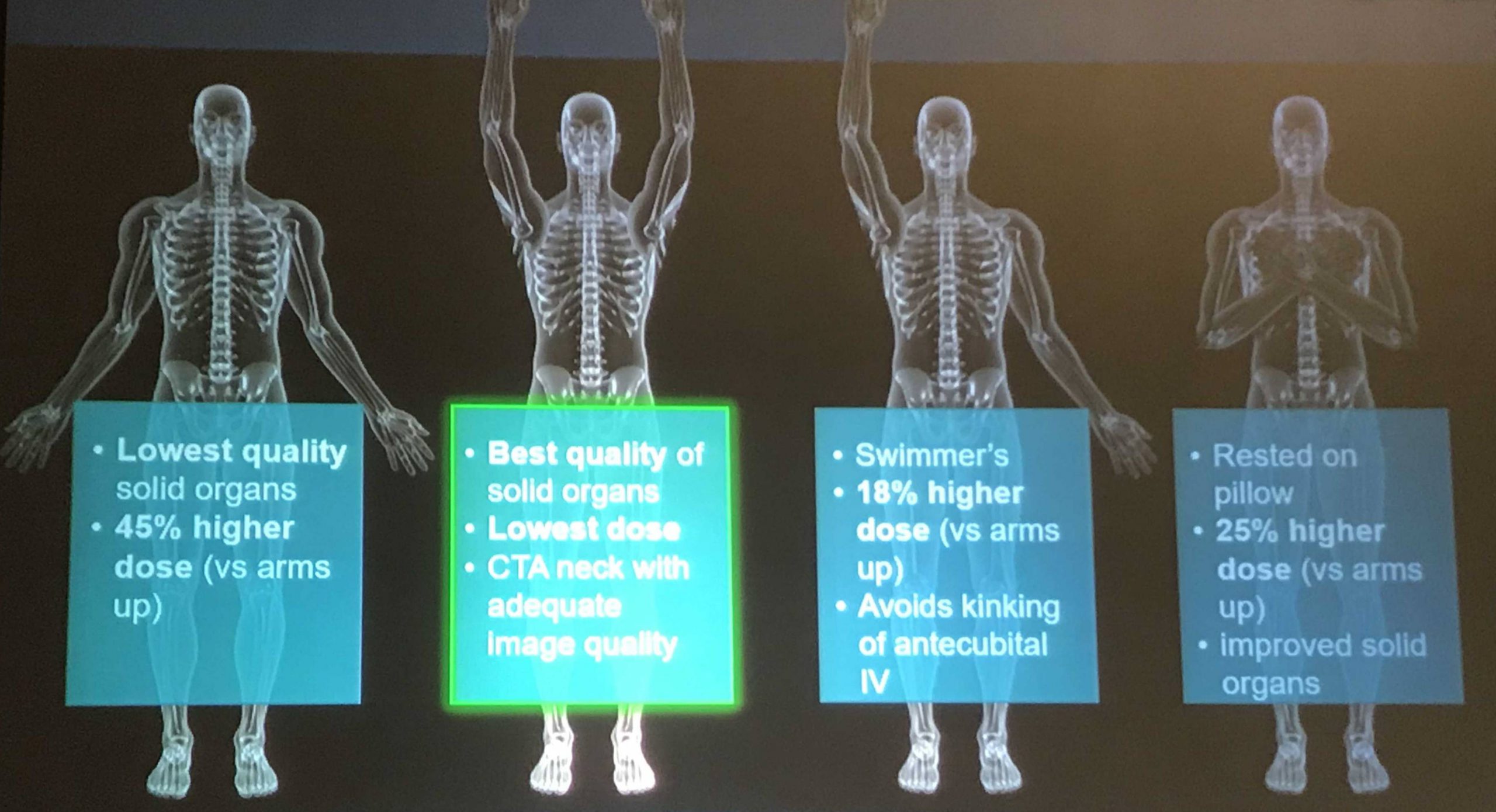
CT scan is a valuable tool for initial screening and diagnosis of trauma patients. However, more attention is being paid to radiation exposure and dosing. Besides selecting patients carefully and striving for ALARA radiation dosing (as low as reasonably achievable) by adjusting technique, what else can be done? Obviously, shielding parts of the body that do not need imaging is simple and effective. But what about simply changing body position?

One simple item to consider is arm positioning in torso scanning. There are no consistent recommendations for use in trauma scanning. Patients with arm and shoulder injuries generally keep the affected upper extremity at their side. Radiologists prefer to have the arms up if possible to reduce scatter and provide clearer imaging.

Radiation physics research has examined arm positioning and its effect on radiation dose. A retrospective review of 690 patients used dose information computed by the CT software and displayed on the console. Radiation exposure was estimated using this data and was stratified by arm positioning. Even though there are some issues with study design, the results were impressive.

The dose results were as follows:

  • Both arms up: 19.2 mSv (p<0.0000001)
  • Left arm up: 22.5 mSv
  • Right arm up: 23.5 mSv
  • Arms down: 24.7 mSv

Bottom line: Do everything you can to reduce radiation exposure:

  1. Be selective with your imaging. Do you really need it?
  2. Work with your radiologists and physicists to use techniques that reduce dose yet retain image quality
  3. Shield everything that’s not being imaged.
  4. Think hard about getting CT scans in children
  5. Raise both arms up during torso scanning unless injuries preclude it.

Reference: Influence of arm positioning on radiation dose for whole body computed tomography in trauma patients. J Trauma 70(4):900-905, 2011.

The Cost Of Duplicate Radiographic Studies

Speaking of radiation, here’s another tidbit. Duplicate radiographic studies are a continuing issue for trauma professionals, particularly after transfer from a smaller hospital to a trauma center. The incidence has been estimated anywhere from 25% to 60% of patients. A lot has been written about the radiation dangers, but what about cost?

A Level II trauma center reviewed their experience with duplicate studies in orthopedic transfer patients retrospectively over a one year period. They looked at the usual demographics, but also included payor, cost information, and reason for repeat imaging. Radiation dose information was also collected.

Here are the factoids:

  • 513 patients were accepted from 36 referring hospitals
  • 48% had at least one study repeated, 256 CT scans and 161 conventional imaging studies
  • Older patients and patients with low GCS were much more likely to receive repeat studies
  • There were no association with the size of the referring hospital or the ability of the patient to pay
  • Most transfers had commercial insurance; only 11% had Medicaid and 17% were uninsured
  • Additional radiation from repeat scans was 8 mSv. The average radiation dose from both hospitals was 38 mSv. This is 13 years of background radiation exposure!
  • The cost of all the repeat studies was over $96,000

Bottom line: This is an eye-opening study, particularly regarding how often repeat imaging is needed, how much additional radiation is delivered, and now, the cost. And remember that these are orthopedic patients, many of whom had isolated bony injuries. I would expect that patients with multiple and multi-system injuries would require more repeat imaging and waste even more money. It is imperative that all centers that receive transfers look at adopting some kind of electronic data transfer for imaging, be it a VPN or some cloud-based service. With the implementation of the Orange Book by the American College of Surgeons, Level I and II centers will receive a deficiency if they do not have some reliable mechanism for this.

“Level I and II facilities must have a mechanism in place to view radiographic imaging from referring hospitals within their catchment area (CD 11–42).”

Reference: Clinical and Economic Impact of Duplicated Radiographic Studies in Trauma Patients Transferred to a Regional Trauma Center. J Ortho Trauma 29(7):e214-e218, 2015.

How Much Radiation is the Trauma Team Really Exposed To?

Previously, I posted about “other people” wearing perfectly good lead aprons lifting them up to their chin during portable xrays in the trauma bay. Is that really necessary, or is it just an urban legend?

Lead apron fly

After hitting the medical radiation physics books (really light reading, I must say), I’ve finally got an answer. Let’s say that the xray is taken in the “usual fashion”:

  • Portable technique in your trauma bay
  • Tube is approximately 5 feet above the xray plate
  • Typical chest settings of 85kVp, 2mAs, 3mm Al filtration
  • Xray plate is 35x43cm

The calculated exposure to the patient is 52 microGrays. Most of the radiation goes through the patient onto the plate. A very small amount reflects off their bones and the table itself. This is the scatter we worry about.

So let’s assume that the closest person to the patient is 3 feet away (1 meter). Remember that radiation intensity diminishes as the square of the distance. So if the distance doubles, the intensity decreases to one fourth. By calculating the intensity of the small amount of scatter at 3 feet from the patient, we come up with a whopping 0.2 microGrays. Since most people are even further away, the dose is much, much less for them.

Let’s put it perspective now. The background radiation we are exposed to every day (from cosmic rays, brick buildings, etc) amounts to about 2400 microGrays per year. So 0.2 microGrays from chest xray scatter is less than the radiation we are exposed to naturally in about 44 minutes!

The bottom line: unless you need to work out you shoulders and pecs, don’t bother to lift your lead apron every time the portable xray unit beeps. It’s a waste of time and effort, unless you are dealing with xray imaging on a very regular basis! And that 52 microGrays the patient absorbed? That’s 8 days worth of background radiation.

Wear The Damn Lead Gown!

Pet peeve time. All trauma team members must wear personal protective equipment (PPE) when they attend a trauma activation. It’s for their own protection as well as our patients’. When I am the primary faculty at any trauma activation, I quickly scan all the other team members to ensure they are wearing it. If not, I give them a “gentle reminder” that they need to go get dressed properly.

This is all well and good. But recently I’ve noticed a trend when it comes time to shoot the basic x-rays needed for assessment (chest and/or pelvic images). When the radiology tech calls out to clear the torso and make sure someone else’s head or hands are not over the patient, half the team goes running out of the room. They are missing one key component of their gear:

Yes, their lead gown! Now granted, the amount of radiation exposure is not huge as I’ve documented in previous posts. But it is cumulative and for safety reasons, x-ray exposure must be limited.

But is running out of room the best way to decrease exposure? I think not! This is very disruptive to the way the team should function and interrupts patient care. Ideally, everyone in the room within 2-3 meters of the x-ray tube should be shielded in some way. And the most effective way to do this is to wear the damn lead gown!

Bottom line: I’ve adjusted my scan when the trauma team assembles. I now look for a lead gown underneath the usual PPEs. And if I don’t see it, I remind the offenders that, if they leave the room when the x-rays are taken, I’m not letting them back in. It’s been very effective at reversing this troubling trend.

In my next two posts, I’ll detail how much radiation the team is exposed to, and how much our patients receive from the studies we order.