Tag Archives: pneumothorax

Pneumomediastinum After Falling Down

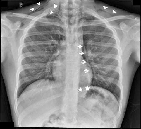
Finding pneumomediastinum on a chest xray or CT scan always gets one’s attention. However, seeing this condition after a simple fall from standing is very simple to evaluate and manage.

There are 3 potential sources of gas in the mediastinum after trauma:

  • Esophagus
  • Trachea
  • Smaller airways / lung parenchyma

Blunt injury to the esophagus is extremely rare, and probably nonexistent after just falling down. Likewise, a tracheal injury from falling over is unheard of. Both of these injuries are far more common with penetrating trauma.

This leaves the lung and smaller airways within it to consider. They are, by far, the most common sources of pneumomediastinum. The most common pattern is that this injury causes a small pneumothorax, which dissects into the mediastinum over time. On occasion, the leak tracks along the visceral pleura and moves directly to the mediastinum.

Management is simple: a repeat chest xray after 6 hours is needed to show non-progression of any pneumothorax, occult or obvious. This image will usually show that the mediastinal air is diminishing as well. There is no need for the patient to be kept NPO or in bed. Monitor any subjective complaints and if all progresses as expected, they can be discharged after a very brief stay.

Tomorrow: A more interesting (and complicated) case of pneumomediastinum.

Flying Or Diving After Traumatic Pneumothorax

Patients who have sustained a traumatic pneumothorax occasionally ask how soon they can fly in an airplane or scuba dive after they are discharged. What’s the right answer?

The basic problem has to do with Boyle’s Law (remember that from high school?). The volume of a gas varies inversely with the barometric pressure. So the lower the pressure, the larger a volume of gas becomes. Most of us hang out pretty close to sea level, so this is not an issue. But for flyers or divers, it may be.

Flying

Helicopters typically fly only one to two thousand feet above the ground, so the air pressure is about the same as standing on the earth. However, flying in a commercial airliner is different. Even though the aircraft may cruise at 30,000+ feet, the inside of the cabin remains considerably lower though not at sea level. Typically, the cabin altitude goes up to about 8,000 to 9,000 feet. Using Boyle’s law, any volume of gas (say, a pneumothorax in your chest), will increase by about a third on a commercial flight.

The physiologic effect of this increase depends upon the patient. If they are young and fit, they may never know anything is happening. But if they are elderly and/or have a limited pulmonary reserve, it may compromise enough lung function to make them symptomatic. And having a medical problem in an aluminum tube at 30,000 feet is never good.

Commercial guidelines for travel after pneumothorax range from 2-6 weeks. The Aerospace Medical Association published guidelines that state that 2-3 weeks is acceptable. The Orlando Regional Medical Center reviewed the literature and devised a practice guideline that has a single Level 2 recommendation that commercial air travel is safe 2 weeks after resolution of the pneumothorax, and that a chest x-ray should be obtained immediately prior to travel to confirm resolution.

Diving

Diving would seem to be pretty safe, right? Any pneumothorax would just shrink while the diver was at depth, then re-expand to the original size when he or she surfaces, right?

Not so fast. You are forgetting why the pneumothorax was there in the first place. The lung was injured, most likely via tearing it, penetration by something sharp, or popping a bleb. If the injured area has not completely healed, then air may begin to escape through it again. And since the air used in scuba diving is delivered under pressure, this could result in a tension pneumothorax.  This is disastrous underwater!

Most injuries leading to pneumothorax heal completely. However, if there are bone spicules stuck in the lung or more complicated parenchymal injuries from penetrating injury, they may never completely heal. This makes the diver susceptible to a tension pneumothorax anytime they use their regulator.

Bottom line: Most patients can safely travel on commercial aircraft 2 weeks after resolution of pneumothorax. Ideally, a chest xray should be obtained shortly before travel to confirm that it is gone. Helicopter travel is okay at any time, since they typically fly at 1,500 feet or less.

Divers should see a physician trained in dive medicine to evaluate their injury and imaging prior to making another dive.

References:

  • Divers Alert Network – Pneumothorax – click to download
  • Practice Guideline, Orlando Regional Medical Center. Air travel following traumatic pneumothorax. October 2009.
  • Medical Guidelines for Airline Travel, 2nd edition. Aerospace Medical Association. Aviation, Space, and Environmental Medicine 74(5) Section II Supplement, May 2003.
When To Remove a Chest Tube

Chest tubes are needed occasionally to help manage chest injuries. How do you decide when they are ready for removal?

Unfortunately, the literature is not very helpful in answering this question. To come up with a uniform way of pulling them, our group looked at any existing literature and then filled in the (many) blanks, negotiating criteria that we could all live with. We came up with the following.

Removal criteria:

  1. No (or a minimal, stable) residual pneumothorax
  2. No air leak
  3. Less than 150cc drainage over the last 3 shifts. We do not use daily volumes, as it may delay the removal sequence. We have moved away from the “only pull tubes on the day shift” mentality. Once the criteria are met, we begin the removal sequence, even in the evening or at night. This typically shaves half a day from the hospital stay.

Removal sequence:

  • Has the patient ever had an air leak? If so, they are placed on water seal for 6 hours and a followup AP or PA view chest x-ray is obtained. If no pneumothorax is seen, proceed to the next step.
  • Pull the tube. See tomorrow’s blog for a video on how to do it.
  • Obtain a followup AP or PA view chest x-ray in 6 hours.
  • If no recurrent pneumothorax, send the patient home! (if appropriate)

Click here to download the full printed protocol.

Flying After Pneumothorax

This question just keeps on coming up!

Patients who have sustained a traumatic pneumothorax occasionally ask how soon they can fly in an airplane after they are discharged. What’s the right answer?

The basic problem has to do with Boyle’s Law (remember that from high school?). The volume of a gas varies inversely with the barometric pressure. So the lower the pressure, the larger a volume of gas becomes. Most of us hang out pretty close to sea level, so this is not an issue.

However, flying in a commercial airliner is different. Even though the aircraft may cruise at 30,000+ feet, the inside of the cabin remains considerably lower though not at sea level. Typically, the cabin altitude goes up to about 8,000 to 9,000 feet. Using Boyle’s law, any volume of gas (say, a pneumothorax in your chest), will increase by about a third on a commercial flight.

The physiologic effect of this increase depends upon the patient. If they are young and fit, they may never know anything is happening. But if they are elderly and/or have a limited pulmonary reserve, it may compromise enough lung function to make them symptomatic.

Commercial guidelines for travel after pneumothorax range from 2-6 weeks. The Aerospace Medical Association published guidelines that state that 2-3 weeks is acceptable. The Orlando Regional Medical Center reviewed the literature and devised a practice guideline that has a single Level 2 recommendation that commercial air travel is safe 2 weeks after resolution of the pneumothorax, and that a chest xray should be obtained immediately prior to travel to confirm resolution.

Bottom line: Patients can safely travel on commercial aircraft 2 weeks after resolution of pneumothorax. Ideally, a chest xray should be obtained shortly before travel to confirm that it is gone. Helicopter travel is okay at any time, since they typically fly at 1,500 feet or less.

References:

  • Practice Guideline, Orlando Regional Medical Center. Air travel following traumatic pneumothorax. October 2009.
  • Medical Guidelines for Airline Travel, 2nd edition. Aerospace Medical Association. Aviation, Space, and Environmental Medicine 74(5) Section II Supplement, May 2003.
What Percent Pneumothorax Is It?

Frequently, radiologists and trauma professionals are coerced into describing the size of a pneumothorax seen on chest xray in percentage terms. They may something like “the patient has a 30% pneumothorax.”

The truth is that one cannot estimate a 3D volume based on a 2D study like a conventional chest xray. Everyone has seen the patient who has no or a minimal pneumothorax on a supine chest xray, only to discover one of significant size with CT scan.

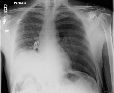
Very few centers have the software that can determine the percentage of chest volume taken up with air. There are only two percentages that can be determined by viewing a regular chest xray: 0% and 100%. Obviously, 0% means no visible pneumothorax, and 100% means complete collapse. Even 100% doesn’t really look like 100% because the completely collapsed lung takes up some space. See the xray at the top for a 100% pneumothorax.

If you line up 10 trauma professionals and show them a chest xray with a pneumothorax, you will get 10 different estimates of their size. And there aren’t any guidelines as to what size demands chest tube insertion and what size can be watched.

The solution is to be as quantitative as possible. Describe the pneumothorax in terms of the maximum distance the edge of the lung is from the inside of the chest wall, and which intercostal space the pneumothorax extends to. So instead of saying “the patient has a 25% pneumo,” say “the pneumothorax is 1 cm wide and extends from the apex to the fifth intercostal space on an upright film.”