Tag Archives: pelvic fracture

Pelvic Fracture Intervention And Venous Thromboembolism Risk

Earlier this year, I wrote a series of posts on the two commonly used pelvic fracture interventions: preperitoneal packing (PPP) and angioembolization (AE). To sum up, both are equally effective in controlling hemorrhage, but the hospital costs for patients undergoing angioembolization are significantly less. This is probably because there is no need to perform repeated operations to insert and remove the preperitoneal packs when angiography is used.

But what about venous thromboembolism risk? Patients with pelvic fractures are already at high risk for it. Couldn’t the increase in pressure on the pelvic veins or the use of thrombogenic materials increase the risk? Authors from several well-known trauma centers collaborated to re-analyze data from the CLOTT study. This is another cute acronym for an extensive study, the Consortium of Leaders in the Study of Traumatic Thromboembolism. It looked at the incidence and risk factors for VTE in trauma patients. This analysis focused on VTE risk in patients with pelvic fractures, comparing those undergoing one of the two interventions with those who had neither.

Here are the factoids:

  • The original data were derived from a 17-center study conducted from 2018-2020; this study only included 1,387 patients who had pelvic fractures (as well as other injuries).
  • The primary outcome was the development of VTE during the hospitalization. DVT was detected using duplex ultrasound, and PE was detected by CT angiography. If pulmonary clots were seen without concomitant DVT, they were not considered to be of embolic origin.
  • For all comers, the overall incidence of VTE was 5.6%
  • Breaking them down by type, there were 2.7% PE, 2.7% DVT, and 1.9% pelvic thrombi (some patients had more than one event)
  • Chemical prophylaxis appeared to be very effective. If started within 24 hours, the incidence was 2.9% vs. 8.4%* if started later.
  • Missed doses did not appear to increase the incidence of VTE (about 5% for both groups)
  • Patients with PPP had a 9% incidence of VTE, pelvic angioembolization had 2.6%, and patients with ORIF had 16%. The incidence was 5.7% if no interventions at all were performed and 16% if more than one occurred.

Bottom line: There are a lot of tidbits in this paper. Most importantly, the use of PPP or AA does not significantly increase VTE risk. Interestingly, ORIF of the pelvis increases it. It’s not clear whether this is due to the procedure itself or is just a surrogate for the severity of pelvic injury. Multivariate analysis suggests that this is not a significant risk factor.

The finding that early chemoprophylaxis reduced VTE incidence to only 3% is very interesting. All too often, prophylaxis is delayed due to solid organ or head injuries. If it can be started safely in such patients, it should be to reduce the occurrence of this complication. One of the banes of management of major trauma is the potential need for repeated surgical procedures, which leads to a phenomenon known as “prophylaxis interruptus.” Thankfully, this study did not find that this increased VTE risk, although it did not stratify by how many doses were missed.

So put your mind at ease about increasing the risk of VTE risk by using procedures to decrease bleeding through mechanical means. But do remember to begin chemoprophylaxis soon and you safely can.

Reference: Does preperitoneal packing increase venous thromboembolism risk among trauma patients? A prospective multicenter analysis across 17 level I trauma centers. Journal of Trauma and Acute Care Surgery 97(5):p 791-798, November 2024.

REBOA For Pelvic Fractures

Resuscitative Endovascular Balloon Occlusion of the Aorta (REBOA) is one of the newer shiny toys that trauma professionals have adopted over the past 8 years or so. It is used to buy time for a patient who is near arrest in order to temporarily stop bleeding and get them to the operating room.

And as with all new toys, everyone wants one! I have always advised caution. Adopt a data-based approach to toy usage. Unfortunately information has been accumulating ever so slowly on this one. To help remedy this, the AAST created a registry in 2013 to consolidate the low REBOA experience numbers accruing across the US.

A group of seven surgeons representing higher-volume REBOA centers collaborated to review the AAST AORTA registry, which prospectively collects data on patients who undergo aortic occlusion. They retrospectively reviewed over six years of data on adult patients receiving REBOA for pelvic injury. They examined demographic, procedural, and outcome data in patients who underwent this procedure, both with and without other interventions like preperitoneal packing, angioembolization, or external fixation. For inclusion in the study, patients needed to have sustained blunt trauma and survived beyond the emergency department.

Here are the factoids:

  • Of the 207 patients with pelvic (Zone 3) REBOA in the registry, only 160 met inclusion criteria
  • Patients who only had REBOA suffered a mortality rate of 40% (5% in OR and 35% in the ICU)
  • Patients who had REBOA plus one of the other interventions had a 31% mortality rate (6% in OR and 25% in ICU)
  • Patients who had REBOA plus two other interventions also had a 31% mortality rate (12% in OR and 30% in the ICU)
  • Adding external fixation with or without another adjunct appeared to decrease mortality by half (from 50% to about 25%)
  • Complications were very common in all subsets, ranging from 35% to 86%
  • Patients receiving more interventions typically were more severely injured
  • No combination of REBOA and adjuncts was superior, but addition of an external fixator did appear to improve survival
  • Patients receiving angiographic embolization had a higher incidence of AKI, sometimes resulting in the need for dialysis
  • There were no significant outcome differences with REBOA use alone or with additional adjuncts

Bottom line: This was a primarily a descriptive study of how REBOA is integrated into pelvic fracture care at select US trauma centers. It was not really designed to compare the efficacy of REBOA vs preperitoneal packing vs angioembolization vs external fixation of the pelvis.

But it does show that survival remains dismal in these patients and the complication rates of REBOA + adjunct use are considerable. The authors correctly conclude that REBOA is being used in the treatment of pelvic fractures, frequently with the addition of other adjuncts. They state that the benefit of more interventions must be balanced against the potential for complications. And finally, they note that there is a need to fill in the evidence base if we are ever to adopt REBOA as a standard of care for select pelvic fractures.

What does this mean to all of you who are thinking of playing with this toy? Proceed with caution! The learning curve is steep. The complication rate is high. The opportunity for mayhem is great. This means that you must proceed deliberately. Get some advanced training with this technique. Use your performance improvement program to impartially critique its use with every deployment. And submit your experience to the national registry so we can all learn from your experience and figure out how to optimize use of this tool.

Reference: Patterns and outcomes of zone 3 REBOA use in the management of severe pelvic fractures: Results from the AAST Aortic Occlusion for Resuscitation in Trauma and Acute Care Surgery database. J Trauma 90(4):659-665, 2021.


Preperitoneal Packing vs Angioembolization For Pelvic Fracture

In my last post, I laid out the various options available for initial management of major pelvic fracture bleeding. Today, I’ll compare two of the newer tools: embolization (AE) and preperitoneal packing (PPP). In the next post, I’ll look at the data available for REBOA.

Interestingly, the use of AE and PPP vary geographically. Angioembolization has been a mainstay in the US for some time, and PPP has been more commonly used in Europe. The use of both is becoming more widespread, and each has its pros and cons.

Angioembolization requires the presence of a special interventional radiology team and a reasonably stable patient. The procedure can take some time, and the IR suite is not really the place to house an unstable patient. Preperitoneal packing requires a reasonably stable pelvis to hold the packs in place for optimal tamponade, which may require application of an external fixator at the time of the procedure.

But is one better than the other? A number of relatively small studies have been performed, which means that it is time to synthesize them and see if some clearer answers can be found. The trauma group in Newcastle, Australia did just this. They performed a systematic search of the literature, analyzing the impact of each procedure on in-hospital mortality.

Here are the factoids:

  • A total of 18 studies met the authors’ inclusion criteria: 6 studies on AE, 9 studies of PPP, and 3 that compared them to each other
  • ISS was significantly higher in the PPP group vs AE (41 vs 36)
  • Average time to OR in the PPP patients was 60 minutes vs 131 minutes to IR in the AE group (statistically significant)
  • A quarter (27%) of the PPP patients did not get adequate hemorrhage control and required AE
  • In-hospital mortality in the PPP papers was 23% vs 32% in the AE research
  • Mortality in the papers that compared AE directly to PPP wasno different

Bottom line: What does this all mean? Is packing “better” than embolization? The simple answer is that we don’t know yet. Due to the way this study was performed, it is not possible to tease out all of the possible confounders. 

We are taught that control of hemorrhage is paramount. The time to definitive management in the AE group was twice that of the PPP patients. This could have a major impact on mortality. Two hours of bleeding can certainly kill. And the lower mortality in the PPP group occurred even though their injury severity was higher.

Many trauma centers have both of these interventions available to choose from. How should we approach their use? Unfortunately the literature is still to scarce to come to strong conclusions. Until we have better research to learn from, I suggest the following:

  • Time is of the essence. Which procedure can you get the fastest? In many cases, this will be preperitoneal packing since it’s just a trip to your trauma OR, which should be ready and waiting. If you have an IR team standing by or available very quickly, you could consider them first.
  • Pay attention to hemodynamic stability. An IR suite is no place for an unstable patient. The resuscitation equipment is not on par with the OR, and one never knows exactly how long the procedure will last.
  • If you have a hybrid room, use it! This is the ideal situation. The surgeon can start the PPP while the orthopedic surgeon applies a fixator. And the radiologist can be preparing to finish it off with a quick squirt as soon as they move away from the groin.
  • The use of one does not rule out the other. If one fails and the patient has increasing fluid and blood requirements move immediately to the other procedure to try to get control.

Reference: Preperitoneal packing versus Angioembolization for the initial management of hemodynamically unstable pelvic fracture – A Systematic Review and Meta-Analysis. J Trauma, publish ahead of print, Jan 4 2022, doi: 10.1097/TA.0000000000003528.


Don’t Have A Pelvic Binder? Make Your Own! (Video)

During the past two posts, I’ve reviewed the various pelvic binders available and how much they cost. But what can you do if you find yourself in a situation where you need a binder but don’t have one?

It’s time to go MacGyver!

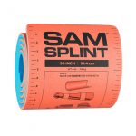
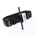
You need three things:

Yes, that’s right. A simple and cheap SAM splint, a tourniquet, and some kind of blade to cut the SAM splint with. Essentially, the SAM splint is the binder and the tourniquet is used to cinch it down in the correct position.

Here’s a video that demonstrates how to do it. Enjoy!

YouTube player

What’s The Best Pelvic Binder? Part 2

Yesterday, I detailed some pelvic binders commonly available in the US. Today, I’ll go through the (little) science there is regarding which are better than others.

And remember, tomorrow I’ll show you how to make a free pelvic binder out of stuff that all medics have in their rig.

There are a number of factors to consider when choosing one of these products. They are:

  • Does it work?
  • Does it hurt or cause skin damage?
  • Is it easy to use?
  • How much does it cost?

It’s difficult to determine how well binders work in the live, clinical setting. But biomechanical studies can serve as a surrogate to try to answer this question. One such cadaver study was carried out in the Netherlands a few years ago. They created one of three different fracture types in pelvis specimens. Special locator wires were placed initially so they could measure bone movement before and after binder placement. All three of the previously discussed commercial binders were used.

Here are the factoids:

  • In fracture patterns that were partially stable or unstable, all binders successfully closed the pelvic ring.
  • None of the binders caused adverse displacements of fracture fragments.
  • Pulling force to achieve complete reduction was lowest with the T-POD (40 Newtons) and highest with the SAM pelvic sling (120 Newtons). The SAM sling limits compression to 150 Newtons, which was more than adequate to close the pelvis.

So what about harm? A healthy volunteer study was used to test each binder for tissue pressure levels. The 80 volunteers were outfitted with a pressure sensing mat around their pelvis, and readings were taken with each binder in place.

Here are the additional factoids:

  • The tissue damage threshold was assumed to be 9.3 kPa sustained for more than 2-3 hours based on the 1994 paper cited below.
  • All binders exceeded the tissue damage threshold at the greater trochanters and sacrum while lying on a backboard. It was highest with the Pelvic Binder and lowest with the SAM sling.
  • Pressures over the trochanters decreased significantly after transfer to a hospital bed, but the Pelvic Binder pressures remained at the tissue damage level.
  • Pressures over the sacrum far exceeded the tissue damage pressure with all binders on a backboard and it remained at or above this level even after transfer to a bed. Once again, the Pelvic Binder pressures were higher. The other splints had similar pressures.

And finally, the price! Although your results may vary due to your buying power, the SAM sling is about $50-$70, the Pelvic Binder $140, and the T-POD $125.

Bottom line: The binder that performed the best (equivalent biomechanical testing, better tissue pressure profile) was the SAM sling. It also happens to be the least expensive, although it takes a little more elbow grease to apply. In my mind, that’s a winning combo. Plus, it’s narrow, which allows easy access to the abdomen and groins for procedures. But remember, whichever one you choose, get them off as soon as possible to avoid skin complications.

References:

  • Comparison of three different pelvic circumferential compression devices: a biomechanical cadaver study. JBJS 93:230-240, 2011.
  • Randomised clinical trial comparing pressure characteristics of pelvic circumferential compression devices in healthy volunteers. Injury 42:1020-1026, 2011.
  • Pressure sores. BMJ 309(6959):853-857, 1994.