Tag Archives: pediatric

Button Batteries: Part 2 – Getting Them Out

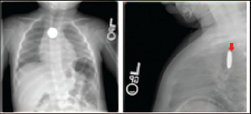
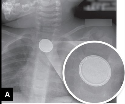
In my last post, I detailed how to suspect and image a button battery ingestion. In this one, I’ll describe how to extract them, and how quickly it’s necessary.

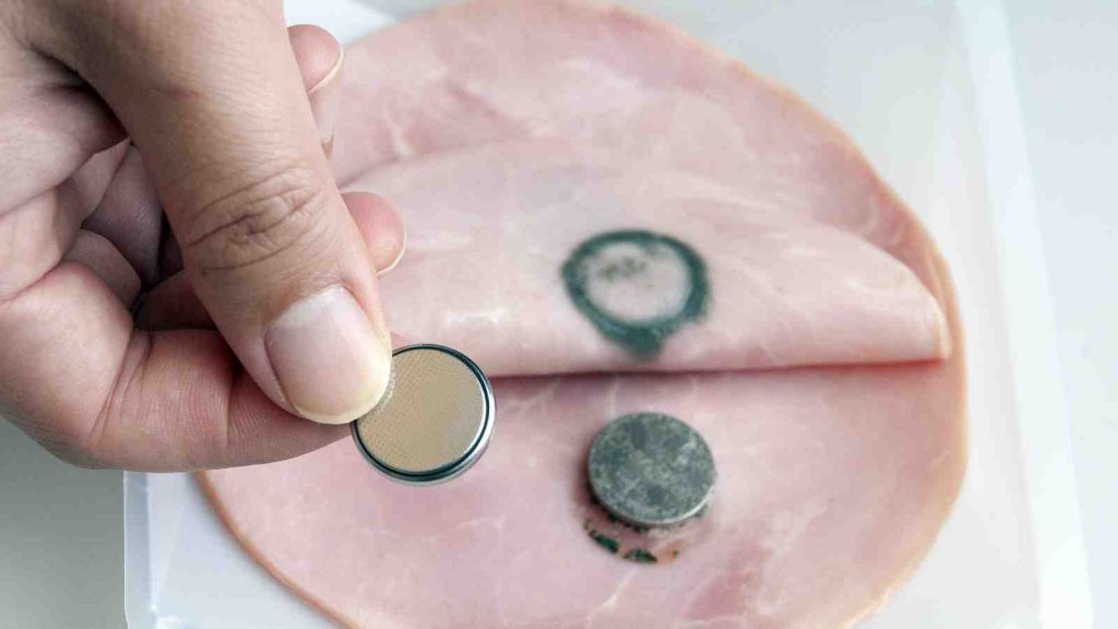
When batteries come to rest and are surrounded by moist mucosal tissue, a current arc is generated around the two sides of the button. This releases heat, which coagulates the surrounding tissue. Depending on the location, closeness of contact, and the duration, these burn injuries may extend into underlying tissue. This is of particular significance in the esophagus, which is in close proximity to the thoracic aorta.

Here’s a simple demonstration you can do at home with some lunch meat:

Here are guidelines for what to do when you encounter pediatric patients who have ingested a button battery:

  • If the child is experiencing bleeding from the upper GI tract, activate your trauma team. The child may have an aorto-esophageal fistula. If there is no active bleeding, obtain a chest x-ray to assess the battery’s position. If there is active bleeding, proceed to the OR (preferably a hybrid room if you have one) and use fluoro to locate the battery. If bleeding persists, call appropriate pediatric surgical specialists (surgery, CV surgery, GI), activate your massive transfusion protocol, and consider tamponade with a Blakemore tube (remember those?) or a urinary catheter if you don’t have one.
  • No bleeding from the upper GI tract? If the battery is large (>20mm) and/or the child is small (<5 years), and is lodged in the esophagus, proceed immediately to OR and remove endoscopically.
  • Batteries in the stomach are of less concern. They will generally pass if <20mm. A repeat x-ray after 48 hours should be obtained for larger batteries. If still in the stomach, they should be removed endoscopically. Smaller batteries will usually pass, and should be re-imaged after two weeks to confirm this.

References:

  • Button battery and magnet ingestions in the pediatric patient.  Curr Opin Pediatrics 30:653-659, 2018.
  • Management of ingested foreign bodies in children: a clinical report of the NASPGHAN Endoscopy Committee. J Pediatric Gastroenterol Nutr 60:562-574, 2015.

Button Batteries: Part 1

I know what you are saying. Button batteries? Trauma? Not too many adult trauma professionals have seen or heard of this. But those who care for pediatric patients should be very familiar. If the importance of this seemingly minor problem is ignored, the results can be catastrophic.

Kids eat stuff, and not just food. The smaller ones always seem to be putting things in their mouths. Foreign body ingestion (or insertion into other orifices) is a common presentation at pediatric emergency departments. Unfortunately, the fact that a battery has been eaten may not be appreciated by the parents.  The child may be brought in with  nonspecific GI or respiratory symptoms.

As soon as a battery ingestion is known or suspected, a two-view chest x-ray is needed. This should show both chest and upper abdomen in order to visualize both esophagus and stomach. Separate chest and abdominal images may be required if the child is too large for a single shot. Two views (AP and lateral) are important because the nature of the foreign body may not be appreciated if the battery is seen edge-on.

If you are fortunate enough to image the battery “face-on”, you may see a telltale halo sign. Because of the way these batteries are put together, there are two metal sides that have a slight difference in overlap.

You’ve made the diagnosis! So now what? And how quickly? I’ll deal with this in my next post.

GCS At 40: Pediatric Glasgow Coma Scale

I’ve been discussing the Glasgow Coma Scale (GCS), but only the adult version so far. The pediatric GCS was created about 10 years after the classic adult scale after it was recognized that several of the scores were not appropriate for younger non-verbal children, typically less than one year of age. It has been validated several times over the ensuing years and has been integrated into our trauma practices.

So what is different about the pediatric GCS scale? It has the same three main components, eye opening, best verbal response, and best motor response. The number of scores under each remains the same as well. The major changes occurred in the verbal response scores. Here’s the breakdown; I’ve highlighted the differences.

Eye Opening

  • All components are the same as for adults

Best Verbal Response

  1. No response to stimuli
  2. Inconsolable, agitated
  3. Inconsistently inconsolable, moaning
  4. Cries but consolable. Has appropriate interactions.
  5. The child smiles, orients to sounds, follows objects, and interacts with adults

Best Motor Response

  1. No response to stimuli
  2. Decerebrate posturing (extension to stimulation, see the adult post for details)
  3. Decorticate posturing (flexion to stimulation, see the adult post for details)
  4. Withdraws from pain
  5. Withdraws from touch
  6. Spontaneous, purposeful movement

In my next post in the series, I’ll review what’s new with the GCS-40 score.

Reference: Neurologic evaluation and support in the child with an acute brain insult. Pediatric Annals 15(1):16-22, 1986.

Radiation Exposure From Imaging At Adult vs Pediatric Trauma Centers

Anyone who reads this blog already knows I am a big believer in well-crafted and focused practice guidelines. And by focused I mean directed toward a clinical problem that typically sees a lot of variability between care providers. Use of imaging is one of these clinical problems. A surgeon may order a certain set of studies for a major blunt trauma patient, and their emergency medicine colleague might order a somewhat different set for someone with the exact same history, physical exam, and injury pattern. Who is right? Neither!

And the variability is even greater when we throw a pediatric patient into the mix. Trauma professionals tend to be even more “generous” when ordering studies on children because they are afraid they might miss something. Unfortunately, this has the potential for overuse of imaging and exposure to unnecessary radiation.

Avery Nathens and a consortium of pediatric trauma centers used the Trauma Quality Improvement Database (TQIP) to review CT imaging practices on children age < 18 over a four year period. Only blunt trauma patients were studied, and the Abbreviated Injury Scale had to be at least 2 for a minimum of one organ system. Transfer patients were excluded because there is no data on imaging for the referring hospital in the TQIP database for them. Comparisons were made between practices at adult trauma centers treating children (ATC), mixed adult/pediatric centers (MTC) and pediatric only trauma centers (PCT).

Here are the factoids:

  • Over 59,000 pediatric trauma patients were identified in the data, and about half (31,081) received at least one CT scan
  • The distribution among the three types of trauma centers was even, with roughly a third seen at each
  • Of the study group 46% had a head CT, 17% a chest CT, and 26% underwent abdominal CT
  • Injured children were more likely to undergo CT if they were older, had a higher ISS, lower motor GCS, were involved in a car crash, or had severe injuries to head or torso
  • Overall CT rates were about the same across the three types of centers (56% ATC, 57% MTC, 43% PTC)
  • Chest CT was performed 8x as much at ATC/MTC vs PTC (!)
  • Abdominal CT was performed 2x as much at ATC/MTC vs PTC
  • Lesser injured children received relatively more CT scans at ATC/MTC when compared to PTC
  • Using standard estimates of cancer risk from all CT scans received, children treated at adult or mixed trauma centers received enough radiation to cause 17 additional lifetime cancers per 100,000 patients
  • About 35 additional lifetime cancers per 100,000 would be caused by the chest and abdominal scans performed at the ATC/MTC centers when compared to pediatric-only centers

Bottom line: This is yet another reason to adopt a well-designed pediatric imaging guideline. Not only are adult centers using CT scanning much more that pediatric-only centers, but they are unnecessarily adding to the lifetime risk for cancer of our children!

As I always recommend, find a well-designed imaging guideline from an established pediatric center and “borrow” it. Sure, it may need a few minor tweaks to fit well with your hospital. That’s okay. Just get it done so your team can begin to order the initial imaging studies consistently and intelligently.

Reference: Computed tomography rates and estimated radiation-associated cancer risk among injured children treated at different trauma center types. Injury 2018, in press.  https://doi.org/10.1016/j.injury.2018.09.036

Phlebotomy And Pediatric Solid Organ Injury

A pediatric trauma paper published a while back tried to focus on reducing the rate of phlebotomy in children who were being observed for solid organ injury. I was more excited about the overall protocol being used to manage liver and spleen injury, as it was a great advance over the original APSA guideline. But let’s look at the phlebotomy part as well.

This is an interestingly weird study, and you’ll see what I mean shortly. Two New York trauma hospitals that take care of pediatric patients pooled 4 years of registry records on children with isolated blunt liver and/or spleen injuries. Then they did a tabletop excercise, looking at “what if” they had applied the APSA guideline, and “what if” they had applied their new, proposed guideline.

Interestingly, this implies that they were using neither! I presume they are trying to justify (and push all their partners) to move to the new protocol from (probably) random, individual choice.

Here are the factoids:

  • 120 records were identified across the 2 hospitals that met criteria
  • Late presentation to the hospital, contrast extravasation, comorbidities, lack of imaging, operative intervention at an outside hospital excluded 59 patients, leaving 61 for analysis. Three of those patients became unstable and were also excluded.
  • None of the remaining patients required operation or angioembolization
  • Use of the “new” (proposed) protocol would reduce ICU admissions by 65%, reduce blood draws by 70%, and reduce hospital stay by 37%
  • Conclusion: use of the protocol would eliminate the need for serial phlebotomy (huh?)

Bottom line: Huh? All this to justify decreasing blood draws? I know, kids hate needles, but the data on decreased length of stay in the hospital and ICU is much more important! We’ve been using a protocol similar to their “new” one at Regions Hospital for almost 10 years, which I’ve shared below. We’ve been enjoying decreased resource utilization, blood draws, and very short lengths of stay for over a decade. And our analysis showed that we save more than $1000 for every patient entering the protocol, compared to the old-fashioned and inefficient way we used to manage them.

In general, kids (and adults) with low grade injuries (I-III) need 2 blood draws, and those with high grade need about 3. Check out our guidelines below to see how it works!

Related posts:

Reference: Reducing scheduled phlebotomy in stable pediatric patients with liver or spleen injury. J Ped Surg 49(5):759-762, 2014.