Tag Archives: fracture

Trauma Mythbusters: NSAIDs And Fracture Healing

Trauma hurts like hell. Over the years, we’ve developed quite a few ways of combating this pain. A number of drug classes have been developed to reduce it. One of the more common non-narcotic drug classes are the NSAIDs. As I’ve mentioned before, every drug has dozens of effects. Drug companies market a particular medication based on one of the predominant effects. All the others are considered side effects.

NSAIDs are not unique; they have lots of side effects as well. In 2003, several papers brought to light possible interactions between these drugs and fracture healing. Specifically, there were questions about these drugs interfering with the healing process and of increasing the number of delayed unions or nonunions. But once again, how convincing were these papers, really?

It would seem to make sense that NSAIDs could interfere with bone healing. This  process relies heavily on the regulation of osteoblast and osteoclast function, which itself is regulated by prostaglandins. Since prostaglandins are synthesized by the COX enzymes, COX inhibitors like the NSAIDs should have the potential to impair this process. Indeed, animal studies in rats and rabbits seem to bear this out.

But as we have seen before, good animal studies don’t always translate well to human experience. Although a study from 2005 suggested that NSAID administration in older patients within 90 days of injury had a higher incidence of fracture nonunion, the study design was not a very good one. It is equally likely that patients who required these drugs in this age group may have been at higher risk for nonunion in the first place.

In fact, there are no large, prospective randomized studies that have explored the effect of short-term or long-term NSAID administration on fracture repair. But there have been several smaller studies that showed absolutely no effect on nonunion with short-term administration of this drug class. Yet the dogma that leads us to avoid giving these drugs persists.

Bottom line: Once again, the animal data is clear but the human data is not. Although there are theoretical concerns about their use, there is not enough solid risk:benefit information to abandon short-term NSAID use in patients who really need them. NSAIDs can and should be prescribed in patients with short-term needs and simple fractures.

References:

  1. Effects of nonsteroidal anti-inflammatory drugs on bone formation and soft-tissue healing. J AM Acad Orthop Surg 12:139-43, 2004.
  2. Effect of COX-2 on fracture-healing in the rat femur. J Bone Joint Surg Am 86:116-123, 2004.
  3. Effects of perioperative anti-inflammatory and immunomodulating therapy on surgical wound healing. Pharmacotherapy 25:1566-1591, 2005.
  4. Pharmacological agents and impairment of fracture healing: what is the evidence? Injury 39:384-394, 2008.
  5. High dose nonsteroidal anti-inflammatory drugs compromise spinal fusion. Can J Anaesth 52:506-512, 2005.

Giving Vitamin D After Fracture

The role of Vitamin D in fracture healing is well known. So, of course, trauma professionals have tried to promote Vitamin D

supplementation to counteract the effects of osteoporosis. A meta-analysis of of 12 papers on the topic relating to hip and other non-vertebral fractures showed that there was roughly a 25% risk reduction for any non-vertebral fractures in patients taking 700-800 U of Vitamin D supplements daily.

Sounds good. So what about taking Vitamin D after a fracture occurs? Seems like it should promote healing, right? A very recent meta-analysis that is awaiting publication looked at this very question.

Unfortunately, there was a tremendous variability in the interventions, outcomes, and measures of variance. All the authors could do was summarize individual papers, and a true meta-analysis could not be performed.

Here are the factoids:

  •  81 papers made the cut for final review
  • A whopping 70% of the population with fractures had low Vitamin D levels
  • Vitamin D supplementation in hospital and after discharge did increase serum levels
  • Only one study, a meeting abstract which has still not seen the light of day in a journal, suggested a trend toward less malunions following a single loading dose of Vitamin D

Bottom line: Vitamin D is a great idea for people who are known to have, or are at risk for, osteoporosis and fractures. It definitely toughens up the bones and lowers the risk of fracture. However, the utility of giving it after a fall has not been shown. Of the 81 papers reviewed, none showed a significant impact on fracture healing. The only good thing is that Vitamin D supplements are cheap. Giving them may make us think that we are helping our patient heal, but it’s not. 

Related posts:

References:  

  • What is the role of vitamin D supplementation in acute fracture patients? A systematic review and meta-analysis of the prevalence of hypovitaminosis D and supplementation efficacy. J Orthopaedic Trauma epub Sep 22 2015.
  • Fracture prevention with vitamin D supplementation: a meta-analysis of randomized controlled trials. JAMA 293(18):2257-2264, 2005.

Displaced Proximal Humerus Fractures – To Operate Or Not?

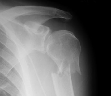
Sometimes you may look at a fracture on an x-ray and cringe. It just seems intuitive that an orthopaedic surgeon should do something about it.

image

But as with many things in the field of medicine, what seems intuitive is not always right. For quite some time, many of these fractures have been managed without surgery, but there has been a more recent trend toward operation. 

Is it the right thing to do? Many patients with this injury are old, with pre-existing medical problems that increase the risk of surgery. A consortium of hospitals in the UK decided to examine this issue with a multi-center trial. They recruited patients from 33 hospitals that could provide either operative or nonoperative management of these fractures.

Here are the factoids:

  • 1250 patients were recruited, and 1000 were excluded. The patients were predominantly elderly (average age 66), and many had comorbidities, insufficient mental capacity, an associated dislocation or clear indication for surgery, or fracture of the other upper extremity.
  • The remaining 250 patients were equally randomized between operative and nonoperative groups.
  • All patients received surgery or a sling, as well as rehabilitation therapy afterwards.
  • After attrition over the 2 year followup period, 114 patients remained in the surgical group and 117 in the nonsurgical group. Overall, they remained well matched.
  • There was no difference in shoulder function or physical well-being between the 2 groups at any time during the 2 years as measured by the Oxford Shoulder Score of the SF-12 Health Survey and the SF-12. Both are self-reported and were administered by a separate examiner.
  • There were 30 complications in the surgical group vs 23 in the nonsurgical group (not significant). One in each group required later surgery.

Bottom line: This is a decent randomized study of a specific clinical question. The large exclusion numbers are a little bothersome, but the authors believed that they had adequate statistical power with their final number of patients. Interestingly, a Cochrane review from 2012 showed similar results with self-reported functional scores, but found a significant number of the nonop patients went on to require surgery. But note that the Cochrane review was an analysis of 6 separate studies, which may weaken their conclusions a bit.

Ultimately, I think that we don’t have any solid conclusions yet. But given the quality of this study, we should start to seriously question whether patients with this fracture, especially elderly ones, really need operative treatment.

Related posts:

Reference: Surgical vs nonsurgical treatment of adults with displaced fractures of the proximal humerus. JAMA 313(10):1037-1047, 2015.

New Technology: 3-D Printed Casts For Fractures

I’ve written quite a lot about the promise of medical applications for 3-D printers. Here’s another one for use by trauma professionals.

Look at the good, old-fashioned plaster cast. It’s been around for decades, and serves its purpose well. It’s easy to apply, inexpensive, and reasonably durable.

Then, along came fiberglass. It’s lighter, more durable, and a bit more water-resistant. And not a whole lot more expensive.

But both of these items have drawbacks. They are heavy. It’s best not to get them wet. Their application is very operator dependent. And probably most importantly, they are opaque. This masks any wounds or skin conditions under it for an extended period of time.

Deniz Karasahin, a Turkish student, won a design award for the development of a 3-D printed cast. It used the appearance of cancellous bone as a model, and is aesthetically very cool. A body scanner is used to scan the affected extremity so that the cast can be customized to the patient. The actual cast is printed from plastic, and can be rendered in a variety of colors. It is hinged, and locks together with a simple pin mechanism.

Bottom line: This is an interesting development in 3-D printing. However, it is not for everybody. Cheap plaster and fiberglass casts are very suitable for many patients. But for some, having the ability to inspect the underlying skin or deal with wounds will make this item much more desirable. And keep in mind, this product was developed for aesthetics. The holes can be much larger and still maintain strength and rigidity. So the cast of the future could be mostly holes, making it very light and shower compatible. Many people might be willing to pay a little more for this convenience.

Note: Ignore the LIPUS ultrasound units that can be incorporated into the one in the article. This is still unproven technology and I don’t recommend it.

Reference / photo credit: A’Design Award Competition

Dysphagia and Cervical Spine Injury

Cervical spine injury presents a host of problems, but one of the least appreciated ones is dysphagia. Many clinicians don’t even think of it, but it is a relatively common problem, especially in the elderly. Swallowing difficulties may arise for several reasons:

  • Prevertebral soft tissue swelling may occur with high cervical spine injuries, leading to changes in the architecture of the posterior pharynx
  • Rigid cervical collars, such as the Miami J and Aspen, and halo vests all force the neck into a neutral position. Elderly patients may have a natural kyphosis, and this change in positioning may interfere with swallowing. Try extending your neck by about 30 degrees and see how much more difficult it is to swallow.
  • Patients with cervical fractures more commonly need a tracheostomy for ventilatory support and/or have a head injury, and these are well known culprits in dysphagia

A study in the Jan 2011 Journal of Trauma outlines the dysphagia problem seen with placement of a halo vest. They studied a series of 79 of their patients who were treated with a halo. A full 66% had problems with their swallowing evaluation. This problem was associated with a significantly longer ICU stay and a somewhat longer overall hospital stay.

Bottom line: Suspect dysphagia in all patients with cervical fractures, especially the elderly. Carry out a formal swallowing evaluation, and adjust the collar or halo if appropriate. 

Reference: Swallowing dysfunction in trauma patients with cervical spine fractures treated with halo-vest fixation. J Trauma 70(1):46-50, 2011.