Tag Archives: EAST2024

Best Of EAST 2024 #9: The Burden Of Transferred TBI Patients

In theory, tiered trauma centers should allow patients with lesser injuries to be treated at lower levels and more severe trauma at higher-level centers. This parallels the resource availability at those centers. In reality, many patients with injuries that seem complex (solid organ, children, and TBI) are transferred due to a “lack of comfort” in taking care of them or the perception that they may deteriorate quickly.

The truth is that many, if not most, of these patients are discharged shortly after transfer to the higher-level center, with minimal intervention. This burdens the trauma system in several ways. First, trauma professionals at the lower-level centers slowly lose their skills and comfort in taking care of these patients. The prehospital system is already plagued with low resources and a shortage of personnel. Using one of the only ambulances in a rural area for transport to a distant center takes it out of the community, potentially putting the area population at risk for delayed care.

The University of Arizona at Tucson group performed another TQIP study to highlight this problem. They performed a four-year retrospective analysis of transfer data in patients with isolated TBI with intracranial hemorrhage. They observed the number of transferred patients who required CT scans, ICP monitoring or craniotomy/craniectomy, length of stay, and mortality.

Here are the factoids:

  • Of the nearly 120,000 patients with isolated TBI at Level I and II centers, 45% were transferred from other centers
  • Most patients had GCS 14-15 on arrival, but 10% had GCS 8
  • CT was performed in 58%, and another repeat CT in 4%
  • Four percent underwent ICP monitoring, and 12.5% had a crani
  • Mortality was 6.5%
  • Median length of stay was two days, with a range of 1-5
  • 18% were discharged within 24 hours, and 39% within 48 hours

The authors concluded that, while half of isolated TBI patients were treated at high-level trauma centers, one-third were discharged home within 48 hours with no intervention other than a CT scan. They recommend systemwide guidelines to improve resource utilization.

Bottom line: This straightforward analysis highlights one of the most significant issues facing trauma systems: unnecessary transfer. For decades, Level I and II centers were convenient and always available for transfer, even if the indications were questionable. Then COVID came along and changed everything.

Now, resources are tight everywhere. EMS is underpaid and under-reimbursed, and personnel are difficult to recruit. Hospital personnel of all types face low staffing levels, making it more stressful to provide the level of care we are accustomed to. Skilled nursing facilities, rehab centers, and other outpatient care settings face the same problems.

This has created a domino effect, where the lower-level centers want to transfer a patient but can’t find a bed at the higher-level centers. When they do, it takes forever to get them transported. Then, the higher-level centers can’t discharge them if they need any level of care other than home care.

There are many pieces to this puzzle, but this abstract clearly outlines one of them. Lower-level centers are transferring some patients who could actually be admitted to them. Several reasons may be given, but it typically boils down to the surgeons or the hospitalists not being “comfortable” with certain patients or worrying that they could deteriorate.

This paper does not tease out what kind of isolated TBI the patients had, and I recommend they do. There is a big difference in patients with a subarachnoid hemorrhage (SAH) vs. those with an epidural. The number of patients with SAH is far greater, and the vast majority can go home after a brief (or no) observation. The likelihood of deterioration in patients not on blood thinners is nearly zero. 

State trauma systems and higher-level trauma centers should work with their Level III and IV partners to adopt consistent practice guidelines and protocols to stratify these patients to identify those at low risk. The higher-level centers should provide education to help their referral partners develop a baseline comfort level with these patients. This is the only way we can begin to realign the levels of trauma centers with the levels of care needed by our patients.

Reference: Endless highways: the burden of transferred traumatic brain injury patients in the United States. EAST 2024, Podium paper #42.

Best Of EAST 2024 #8: Whole Blood And VTE

The pendulum has swung from the use of whole blood in the early 20th century, to component therapy in the 1960s, and now a gradual move toward incorporating whole blood again. More and more papers are being published, and many trauma centers are looking for ways to integrate whole blood into their massive transfusion protocols.

Much of the literature has been dedicated to safety and effectiveness, but little has examined thrombotic complications from its use.  The trauma group at the University of Texas in Houston performed what looks to be a retrospective review of whole blood usage at two Level I trauma centers. Adult patients receiving at least one emergency-release whole blood unit were compared with those receiving only component therapy. They looked at the incidence of venous thromboembolic (VTE) complications such as pulmonary embolism (PE) or deep venous thrombosis (DVT).

Here are the factoids:

  • Nearly 3,500 patients were enrolled and were fairly evenly split between whole blood and component therapy only
  • Whole blood patients were slightly younger, were much more likely to have penetrating injury, and had significantly higher ISS (26 vs 19)
  • The whole blood patients were also significantly more likely to receive TXA, VTE chemoprophylaxis within 48 hours (86% vs. 79%) and lower 30-day survival (74% vs 84%)
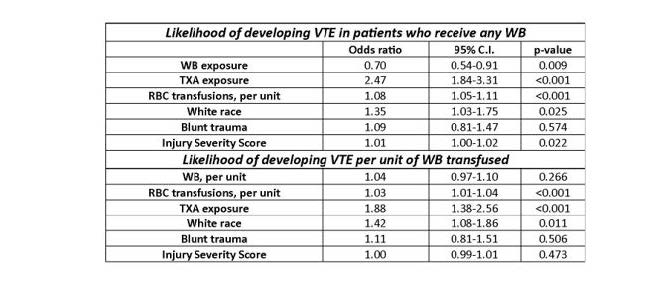
  • Crude incidence of VTE was similar (7% whole blood vs. 9% component), but logistic regression “revealed that whole blood was protective of VTE,” while red cell transfusion and TXA increased VTE risk
  • Each unit of red cells increased VTE risk by 3%

The authors concluded that whole blood was associated with a 30% reduction in VTE, and TX was associated with a 2.5x increase in risk. They cautioned against the use of TXA in the setting of whole-blood resuscitation.

Bottom line: A lot is going on here. First, this is a retrospective study, which limits the number of variables that can be collected reliably. It also makes it much more difficult to perform regression analysis because there are many other possible variables to control for than just the ones collected. 

Next, as quoted in bullet point 4 above, this study can’t show that whole blood was protective, only that it was (maybe) associated with decreased VTE when the variables they collected were controlled. 

Most of the confidence intervals for the “significant” results were very close to the 1.0 line. This leaves the possibility that the result could easily be changed by adding other pertinent variables not included in the data. The only impressive one was the association of TXA exposure and VTE. I think this demands further work.

The authors need to answer several questions in their presentation to help explain the results:

  • Was there any relationship between the number of units of packed cells given and the likelihood of VTE?
  • Similarly, was there a relationship between the number of units of whole blood and possible “protection” from VTE?
  • Did you examine other physiologic or anatomic variables and their relationship with VTE? Specific ones that come to mind are shock, long bone or spine fractures, and TBI. These are some of the variables that need to be included in the regression model to improve it.

Overall, this is an interesting abstract that makes one think. But it either needs some good explanations during the presentation or additional data analysis to make it even more interesting.

Reference: Does whole blood resuscitation increase risk for venous thromboembolism in trauma patients? A comparison of component therapy vs whole blood in 3468 patients. EAST 2024, Podium paper 33.

Best Of EAST #7: King vs. I-Gel Supraglottic Airways

Airway assessment and protection are of paramount importance during trauma care. Airway management is even more challenging in the prehospital environment, where lighting and patient positioning may be suboptimal, and injuries or policies may prohibit orotracheal intubation. A variety of devices have been developed to make airway control simpler and faster.

Both the King and i-gel airways were introduced around 2005. The former functions as an extraglottic airway, and the latter as a supraglottic one. Prehospital providers typically use these devices for airway control across the US. They are both designed for blind insertion without the need for other equipment, such as a laryngoscope.

The group at West Virginia University compared these two devices based on the incidence of hypoxia, cardiac arrest, successful insertion, and survival to hospital discharge. They performed a large database search and attempted to control for patient age, weight, pre-airway hypoxia, and the use of suction.

Here are the factoids:

  • A total of 1,557 patients were studied; one-third received a King airway, and two-thirds had i-gel insertion
  • Half of all patients experienced hypoxia, and a quarter experienced severe hypoxia (saturation < 80%) after insertion of any tube
  • But i-gel placement was not associated with hypoxia, severe hypoxia, cardiac arrest, or decreased survival to discharge, and had better success on first-pass placement

Bottom line: What? This is a first. I honestly can’t figure this abstract out. The two bullets in red above cancel each other out. If half of all patients had hypoxic episodes, and only one-third had King airways, that means that at least 16% of the i-gel patients experienced hypoxia. I can’t reconcile that with the last bullet, where i-gel was not associated with any adverse events.

Several other papers have compared the use of these two devices over the last two decades. Most suggest that the i-gel is simpler, with fewer misplacements and other complications, and tends to be preferred by prehospital personnel.

Unfortunately, I have to disregard this entire abstract due to the conflicting data listed. Perhaps the presenter will clarify or provide some corrections to the data. Otherwise, I have not learned anything from it, and it doesn’t appear to add anything new to the trauma literature.

Reference: A retrospective comparison of the King laryngeal tube and i-gel supraglottic airway devices for injured patients. EAST 2024 Podium paper #27.

Best Of EAST 2024 #6: Pan-Scans In the Elderly

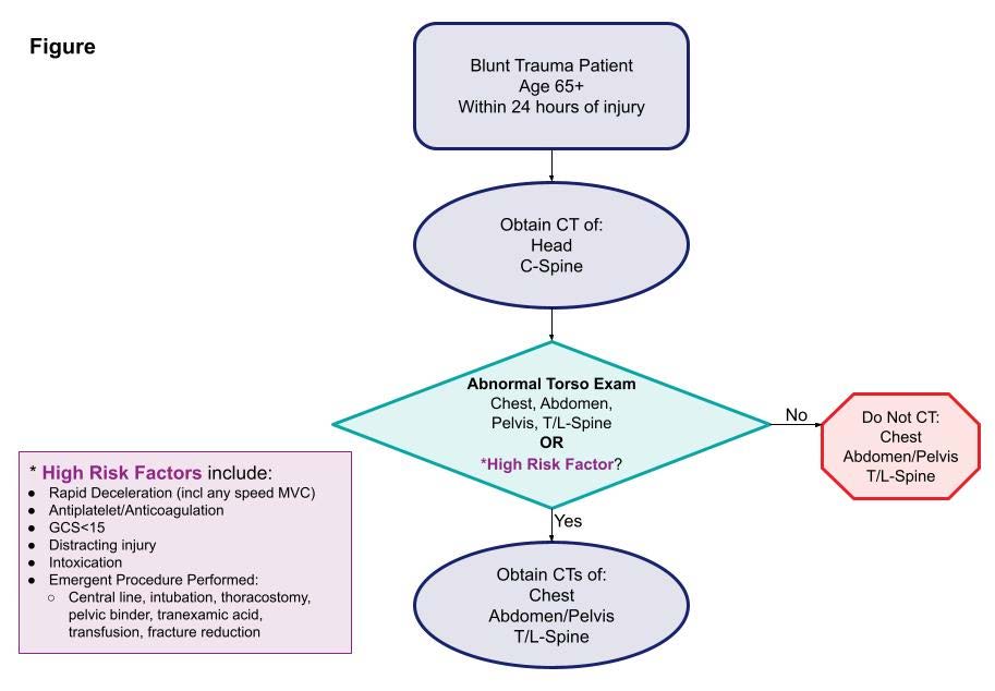
Injured older adults typically sustain those injuries from blunt mechanisms. Radiographic evaluation, particularly CT scans, does not have good supporting literature to dictate which exams should be used in particular patients. There is a long-standing debate on the merits of pan-scan vs. selective scans when using CT.

EAST sponsored a multicenter study to look for specific history and physical exam findings that could help direct CT evaluation. Eighteen Level I and II trauma centers participated in a prospective study of patients aged 65 and above. The authors used machine learning to determine clusters of findings that could be used to create decision rules for using a pan-scan vs a tiered scan (head + cervical spine, then possibly torso). The focus was on injuries to the head/cervical spine and the torso. Patients who presented late after injury (24 hours) or died were excluded, leaving 2,587 for study.

Here are the factoids:

  • The learning system could not develop a rule for pan-scan usage
  • A high-quality model was created to guide the use of selective scanning, which was 94% sensitive with an 86% negative predictive value

  • The authors estimated that 12% of patients would be spared a torso scan using the decision tool

The authors concluded that their decision tool has promising sensitivity and negative predictive value and needs further prospective evaluation to validate it.

Bottom line: As our use of machine learning and AI advances, work like this will continue to accelerate. One of the benefits of using AI as a tool is its ability to sift through enormous amounts of raw data to detect faint but significant signals. 

The downside is that much of what we record is garbage,  making detecting that signal much more difficult. However, the process is inexpensive and can potentially advance health care in a variety of ways. 

The presenter and authors should describe the specific machine learning technique used and outline its particular strengths and weaknesses. Expect to see more and more abstracts like this coming down the pike. And when someone starts applying large language models like Chat-GPT to this type of medical data, look out!

Reference: Scanning the aged to minimize missed injury: an EAST multicenter study. EAST 2024, Podium paper #24.

Best Of EAST #5: How Good Is Lung Ultrasound For Pneumothorax?

Focused abdominal sonography for trauma (FAST) has been a mainstay of rapid diagnosis for many years. The extended FAST exam (eFAST) adds an examination of the thoracic cavities to the basic exam. The sensitivity and specificity of FAST have mostly been determined. However, there is much less literature outlining the accuracy of the eFAST.

The group at Vanderbilt performed a prospective, observational study on the ability of the eFAST exam to detect pneumothorax specifically.  Imaging was performed by a licensed sonographer and read by the attending surgeon and a radiologist as it was completed.

Here are the factoids:

  • Only patients receiving chest X-rays, chest ultrasound, and chest CT for confirmation were included in the study, which totaled 1,499
  • The statistical analysis was as follows:
  • Only 25% of patients had a chest tube placed
  • Patients with false negative exams were more likely to have rib fractures and lower oxygen saturation and blood pressure

The authors concluded that chest ultrasound had a low sensitivity and high false negative rate and that many of the negatives eventually required chest tube insertion. They advise that the chest ultrasound should be used alone with caution.

Bottom line: This is an interesting and relatively large study. However, it is at odds with a paper published in 2021 from George Washington University. They published a series of 3,410 patients and found 71% sensitivity, 99% specificity, 87% PPV, and a 2.2% false negative rate. 

These are very disparate numbers. However, the GW study was retrospective and included both pneumothorax and fluid in the abdomen or chest, whereas this abstract looked strictly at pneumothorax and was prospective.

Both studies are interesting on their own, and the presenter of this abstract will need to explain why the results are so different.  The answer to the question of how much we believe the eFAST result remains up in the air!

References:

  1. Lung ultrasound underdiagnoses clinically significant pneumothorax. EAST 2024, Podium paper 21.
  2. eFAST exam errors at a level 1 trauma center: A retrospective cohort study. Am J Emerg Med. 2021 Nov;49:393-398.