Tag Archives: DVT

Enoxaparin And Pregnancy

Lovenox

Pregnant women get seriously injured, too. And pregnancy is an independent risk factor for deep venous thrombosis. We reflexively start at-risk patients on prophylactic agents for DVT, the most common being enoxaparin. But is it safe to give enoxaparin during pregnancy?

Studies have looked at drug levels in cord blood when the mother is receiving enoxaparin, and none has been found. No specific bleeding complications have been identified, either. So from the baby’s standpoint, administration is probably safe.

However, there are two other issues to consider. In a study looking at the use of enoxaparin for prophylaxis in women with a mechanical heart valve, 2 of 8 women (and their babies) died. Both suffered from clots that developed and blocked the valves. Most likely, the standard dose of enoxaparin was insufficient, so monitoring of anti-Factor Xa levels must be done.

The other problem lies in the multi-dose vial of Lovenox (Sanofi-Aventis). Each 100mg vial contains 45mg of benzyl alcohol, which has been associated with a fatal “gasping syndrome” in premature infants. The individual dose syringes do not have this preservative.

Bottom line: It is probably safe to give enoxaparin to pregnant women after trauma. However, it is unclear if the dose needs to be increased to achieve adequate prophylaxis. Only consider using this medication after consultation with the patient’s obstetrician, and use only the individual dose syringes. Otherwise fall back to standard subcutaneous non-fractionated heparin (even though it is a Category C drug by FDA; it is still considered the anticoagulant of choice during pregnancy).

DVT Prophylaxis After Solid Organ Injury

Nonoperative management of solid organ injury is the norm, and has reduced the operative rate significantly. At the same time, the recognition that development of deep venous thrombosis (DVT) in trauma patients is commonplace creates uncertainty? Is it safe to give chemical prophylaxis with low molecular weight heparin (LMWH)? How soon after injury?

The trauma group at USC+LAC published the findings of a retrospective review of 312 patients undergoing nonoperative management for their liver, spleen or kidney injuries. They looked at chemical prophylaxis administration and its relationship to failure of nonop management of solid organ injury.

As expected, as the grade of the solid organ injury increased, so did the failure rate of nonoperative management. Administration of low molecular weight heparin, such as enoxaparin, did not increase failure rate in this study. All but one failure occurred in patients who had not yet received the injections. Likewise, two DVT and two pulmonary embolisms occurred, but only in patients who had not yet received prophylaxis. 

Bottom line: This small study offers some assurance that early prophylaxis is okay, and a few prospective studies do exist. UCSF / San Francisco General is comfortable beginning chemical prophylaxis 36 hours postop, regardless of solid organ injury. Look for more guidance on this issue in the near future. Until then, consider starting LMWH prophylaxis early to avoid complications from DVT or PE.

Reference: Thromboembolic prophylaxis with low-molecular-weight heparin in patients with blunt solid abdominal organ injuries undergoing nonoperative management: current practice and outcomes. J Trauma 70(1): 141-147, 2011.

DVT In Children

Deep venous thrombosis has been a problem in adult trauma patients for some time. Turns out, it’s a problem in injured children as well although much less common (<1%). However, the subset of kids admitted to the ICU for trauma have a much higher rate if not given prophylaxis (approx. 6%). Most trauma centers have protocols for chemical prophylaxis of adult patients, but not many have similar protocols for children.

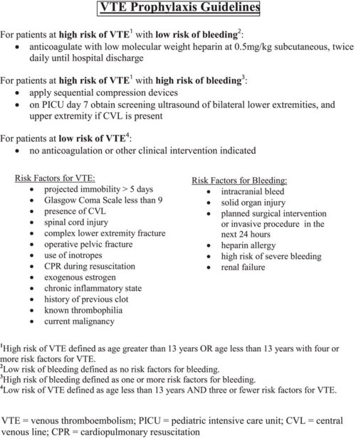
The Medical College of Wisconsin looked at trends prior to and after implementation of a DVT protocol for patients < 19 years old. They used the following protocol to assess risk in patients admitted to the PICU and to determine what type of prophylaxis was warranted:

The need for and type of prophylaxis was balanced against the risk for significant bleeding, and this was accounted for in the protocol. The following significant findings were noted:

  • The overall incidence of DVT decreased significantly (65%) after the protocol was introduced, from 5.2% to 1.8%
  • The 1.8% incidence after protocol use is still higher than most other non-trauma pediatric populations 
  • After the protocol was used, all DVT was detected via screening. Suspicion based on clinical findings (edema, pain) only occurred pre-implementation.
  • Use of the protocol did not increase use of anticoagulation, it standardized management in pediatric patients

Bottom line: DVT does occur in injured children, particularly in severely injured ones who require admission to the ICU. Implementation of a regimented system of monitoring and prophylaxis decreases the overall DVT rate and standardizes care in this group of patients. This is another example of how the use of a well thought out protocol can benefit our patients and provide a more uniform way of managing them.

Related posts:

Reference: Effectiveness of clinical guidelines for deep vein thrombosis prophylaxis in reducing the incidence of venous thromboembolism in critically ill children after trauma. J Trauma 72(5):1292-1297, 2012.

Best Of: Pulmonary Embolism and DVT in Trauma

We have long assumed that pulmonary emboli start as clots in the deep veins of the legs (or pelvis), then break off and float into the branches of the pulmonary artery in the lungs. A huge industry has developed around how best to deal with or prevent this problem, including mechanical devices (sequential compression devices), chemical prophylaxis (heparin products), and physical devices (IVC filters).

The really interesting thing is that less than half of patients who are diagnosed with a pulmonary embolism have identifiable clots in their leg veins. In one study, 26 of 200 patients developed DVT and 4 had a PE. However, none of the DVT patients developed an embolism, and none of the embolism patients had a DVT! How can this kind of disparity be explained?

Researchers at the Massachusetts General Hospital retrospectively looked at the correlation between DVT and PE in trauma patients over a 3 year period. DVT was screened for on a weekly basis by duplex venous ultrsonagraphy. PE was diagnoses exclusively using CT scan of the chest, but also included the pelvic and leg veins to look for a source. A total of 247 patients underwent the CT study for PE and were included in the study.

Forty six patients had PE (39% central, 61% peripheral pulmonary arterial branches) and 18 had DVT (16 seen on the PE CT and 2 found by duplex). Of the 46 patients with PE, only 15% had DVT. All patient groups were similar with respect to injuries, injury severity, sex, anticoagulation and lengths of stay. Interestingly, 71% of PE patients with DVT had a central PE, but only 33% of patients without DVT had a central PE.

The authors propose 4 possible explanations for their findings:

  1. The diagnostics tools for detecting DVT are not very good. FALSE: CT evaluation is probably the “gold standard”, since venography has long since been abandoned
  2. Many clots originate in the upper extremities. FALSE: most centers do not detect many DVTs in the arms
  3. Leg clots do not break off to throw a PE, they dislodge cleanly and completely. FALSE: cadaver studies have not show this to be true
  4. Some clots may form on their own in the pulmonary artery due to endothelial inflammation or other unknown mechanisms. POSSIBLE

An invited critique scrutinizes the study’s use of diagnostics and the lack of hard evidence of clot formation in the lungs.

The bottom line: this is a very intriguing study that questions our assumptions about deep venous thrombosis and pulmonary embolism. More work will be done on this question, and I think the result will be a radical change in our use of anticoagulation and IVC filters over the next 3-5 years.

Reference: Velmahos, Spaniolas, Tabbara et al. Arch Surg. 2009; 144(10):928-932.

DVT: Does spinal cord level make a difference?

Deep venous thrombosis (DVT) is always a concern in trauma patients. Patients with spine and spinal cord injury have been shown to be at higher risk for DVT than many other trauma patients, with a reported incidence ranging from 5% to 70%. However, a few studies have suggested that paraplegics are actually at higher risk than quadriplegics. This just doesn’t seem to make sense.

A NTDB study was done to look at this issue. A total of 18,000+ patients were reviewed, and correlations with spinal cord injury level, demographics, comorbidities and associated injuries were determined.

High cervical (C1-4) and lumbar cord injuries had the lowest DVT rates at about 3%. Lower cervical (C5-7) and high thoracic (T1-6) had the highest rates at 5% and 6.3%, respectively. The lower thoracic spine was about 4.5%. These differences were statistically significant, and the authors also confirmed the usual DVT suspects as being significant (increasing age, increasing injury severity, TBI, chest trauma, and male gender).

Bottom line: Yes, this study does confirm the suspicion that paraplegics are at higher risk for DVT than quadriplegics. Why? We don’t know. And although it is statistically significant, is it clinically significant? I’m not so sure. We’re talking another 1-2 spinal cord injured patients with DVT for every 100 quadriplegics treated. How many do you admit per year? At my institution, this means that there will be 1 additional DVT in this patient group every three to four years. It’s hard to justify making any changes to existing protocols based on these new facts. Always look at the practical side of what you read!

Related posts:

Reference: Risk of venous thromboembolism after spinal cord injury: not all levels are the same. J Trauma 71(5):1241-1245, 2011.