Tag Archives: ct scan

The Role Of Postop CT Scan In Penetrating Trauma

CT scans are commonly used to aid the workup of patients with blunt trauma. They are occasionally useful in penetrating trauma, specifically when penetration into a body cavity is uncertain, and the patient has no hard signs that would send him or her immediately to the operating room.

Is there any role for CT in operative penetrating trauma, after the patient has already been to the OR? The dogma has always been that the eyeballs of the surgeon in the OR are better than any other imaging modality. Really? The surgical group at San Francisco General addressed this question by retrospectively reviewing 6 years of their operative penetrating injury registry data. They were interested in finding how many occult injuries (seen with CT but not by the surgeon) were found on a postop CT. A total of 225 patients who underwent operative management of penetrating abdomen or chest injury were included. Here are the factoids:

  • Only 110 patients had a postop CT scan; 73 had scans within the first 24 hours, the other 37 were scanned later
  • The rationale for early scan was to investigate retroperitoneal injury in half of patients, but frequently no indication was given (41%)
  • The rationale for late scan was for workup of ileus in one-third or for evaluation of new or unexpected clinical problems
  • Occult injuries were found in about half of early CT patients (52%) and 22% of late CT patients
  • The most common occult injuries were fractures, GU issues, regraded solid organ injury, and unrecognized vascular injuries
  • Ten patients had management changes, including:
    • Interventional radiology for four injuries with extravasation
    • Operation for orthopedic or GU injury in seven patients
    • One patient underwent surgery for an unstable spine fracture

Bottom line: There appears to be a significant benefit to sending some penetrating injury patients to CT in the early postop period. Specifically, those with injury to the retroperitoneum, deep into the liver, near the spine, or with multiple and complicated injuries would benefit. Simple stabs and gunshots that stay away from these areas/structures probably do not need follow-up imaging. 

Reference: Routine computed tomography after recent operative exploration for penetrating trauma: What injuries do we miss? J Trauma 83(4):575-578, 2017.

Best Of EAST 2024 #6: Pan-Scans In the Elderly

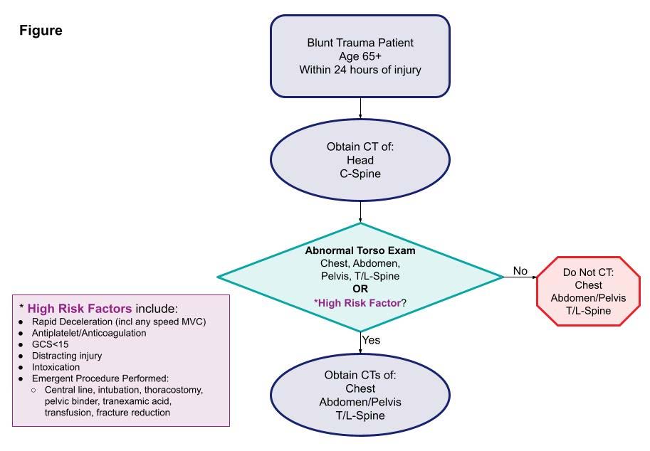
Injured older adults typically sustain those injuries from blunt mechanisms. Radiographic evaluation, particularly CT scans, does not have good supporting literature to dictate which exams should be used in particular patients. There is a long-standing debate on the merits of pan-scan vs. selective scans when using CT.

EAST sponsored a multicenter study to look for specific history and physical exam findings that could help direct CT evaluation. Eighteen Level I and II trauma centers participated in a prospective study of patients aged 65 and above. The authors used machine learning to determine clusters of findings that could be used to create decision rules for using a pan-scan vs a tiered scan (head + cervical spine, then possibly torso). The focus was on injuries to the head/cervical spine and the torso. Patients who presented late after injury (24 hours) or died were excluded, leaving 2,587 for study.

Here are the factoids:

  • The learning system could not develop a rule for pan-scan usage
  • A high-quality model was created to guide the use of selective scanning, which was 94% sensitive with an 86% negative predictive value

  • The authors estimated that 12% of patients would be spared a torso scan using the decision tool

The authors concluded that their decision tool has promising sensitivity and negative predictive value and needs further prospective evaluation to validate it.

Bottom line: As our use of machine learning and AI advances, work like this will continue to accelerate. One of the benefits of using AI as a tool is its ability to sift through enormous amounts of raw data to detect faint but significant signals. 

The downside is that much of what we record is garbage,  making detecting that signal much more difficult. However, the process is inexpensive and can potentially advance health care in a variety of ways. 

The presenter and authors should describe the specific machine learning technique used and outline its particular strengths and weaknesses. Expect to see more and more abstracts like this coming down the pike. And when someone starts applying large language models like Chat-GPT to this type of medical data, look out!

Reference: Scanning the aged to minimize missed injury: an EAST multicenter study. EAST 2024, Podium paper #24.

Best Of EAST 2023 #7: The CT Autopsy

Back in the day, autopsy after trauma death was fairly commonplace. Nowadays, it is typically reserved for fatalities that involve a potential crime. And it can be challenging to get the medical examiner to release copies for trauma performance improvement.

One potential remedy for this began to surface in the literature about twenty years ago: the virtual (or CT) autopsy. This entails sending the postmortem patient to the scanner for head, cervical, chest, and pelvic scans. Although it seems like an exciting idea, there are several logistical issues that I will discuss later.

The trauma group at Indiana University performed a retrospective study to determine the common injury patterns in patients who died at or up to one hour after ED arrival. Their goal was to identify injury patterns that might improve the focus and quality of resuscitative efforts in living patients. They reviewed their experience with doing postmortem CT over a nine-year period. The primary goal was to identify sources of hemorrhage, TBI, and cervical spine injury. They also wanted to identify significant pneumothorax and misplaced airway devices.

Here are the factoids:

  • There were 80 decedents in the study, and they were severely injured, with an average ISS of 42
  • About three quarters arrested prior to arrival, and the remainder arrived with a pulse
  • The most common major injuries were severe TBI (41%), long bone fractures (25%), hemoperitoneum (23%), and cervical spine injury (19%)
  • A moderate pneumothorax was present in 19% of cases
  • Misplaced airway was identified in 5%
  • There was no difference in injury or device mishap patterns between pre-hospital and in-hospital arrest patients (although the number of patients was probably too small to detect one)

The authors concluded that the injury patterns between those who died prior to arrival vs. after were the same. They also noted that patients in arrest should automatically have their chest decompressed and the airway position checked.

Bottom line: This is an intriguing study of a concept I’ve been thinking about for years. The quality improvement benefits could be amazing! Imagine getting immediate feedback on the cause of death and how it might influence future resuscitations. The authors pointed out the power of this with their discovery of missed pneumothorax and malpositioned airways.

But, as mentioned above, there are a host of logistical problems to work out first. Here is a partial list:

  • Who accompanies the patient to scan? A nurse? The team?
  • Covered or uncovered? It might be creepy for people in the hallways to see a covered person being wheeled around. That’s why hospitals always have those white, wheeled boxes. But it’s equally creepy to see a person who is not moving or breathing being transported.
  • Be prepared for your radiologists to gripe about doing free reads
  • Where does the report go? It shouldn’t go to the medical record. Or should it?
  • What about liability issues? If the team misses something big and the report goes to the chart, it’s fair game for a lawsuit.
  • And many more!

Here are my questions and comments for the presenter/authors:

  • How did you come to do this study? It appears that your group has been performing CT autopsies for almost a decade. Was there a protocol? Was it done on every eligible patient? If not, could this have skewed your results?
  • Do you have the statistical power to detect any differences between the various groups? A few of your results did approach significance. Perhaps more subjects would have helped.
  • Tell us how you have addressed the logistical problems above.

This is great work; perhaps it will stimulate a move toward embracing this concept!

Reference: CHARACTERIZATION OF FATAL BLUNT INJURIES USING POST-MORTEM COMPUTED TOMOGRAPHY. EAST 2023 Podium paper #14.


Best of AAST 2022 #4: The “Hybrid ER” – Again?

Two years ago, an abstract was presented at this meeting describing the concept of the “hybrid ER.” Check it out using this link. This concept was pioneered in Japan, and consists of a special trauma resuscitation room in the ED with everything but an operating room built into it. It’s possible to perform whole-body CT scan, interventional procedures, and REBOA without moving the patient. Here’s a picture from that abstract:
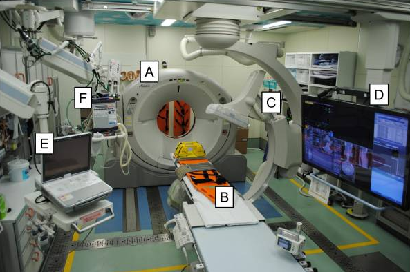
A = CT scanner   B = CT exam table   C = movable C-arm   D = monitor screen   E = ultrasound   F = ventilator

In that abstract, about a thousand patients were compared with two thirds in the hybrid ER group and one third undergoing conventional evaluation. The authors concluded that mortality was significantly improved in the hybrid ER group, and even more so in high ISS patients.

I had a lot of questions for that abstract that were answered in the subsequently published manuscript (reference 2). The authors have updated their experience using new data from the last five years. They created a new approach to resuscitation that is different than the usual ATLS sequence for select patients. Here’s the algorithm they used:
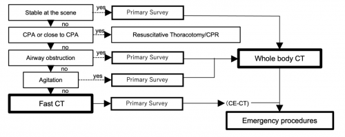
The primary survey is completed, then the patient undergoes a quick whole body CT scan. After that, the secondary survey progresses and any necessary emergency procedures are performed.

In this abstract, the authors compared a group of 46 patients who underwent standard ATLS evaluation with 49 who received the expedited process, which they termed CT First Resuscitation (CTFR). All patients had presumptive hemorrhagic shock based on prehospital vital signs. The authors analyzed injury patterns, interventions performed, timing, adverse events, and outcome. Demographics and injury severity were similar in the two groups.

Here are the factoids:

  • Time to CT in the CTFR group was significantly shorter (1.5 min vs 15 min)
  • The expedited scan settings for CTFR resulted in blindingly fast scan times (median 56 seconds)
  • None of the CTFR patients decompensated during the scan process
  • There was no difference in mortality between CTFR and standard evaluation (14% vs 4%, p=.1)
  • There was no difference in time to hemostatic intervention (56 vs 59 minutes)
  • There was no difference in red cell transfusions (no units in either group)

The authors concluded that CTFR expedited trauma management without adverse effects, and there was no increase in mortality. They, or course, recommended further study.

Bottom line: Several trauma surgeons from a variety of centers wrote an invited commentary last year (reference 3) expressing their excitement about this concept. Reducing time to definitive control of hemorrhage has been repeatedly shown to improve survival. The hybrid ER is one way of reducing those times by eliminating most of the time needed to move the patient about and providing everything but an operating room in the emergency department.

But they also recognized the limitations of this concept. The changes to the ED physical plant are extreme and involve the installation of very expensive equipment that must be heavily shielded from the rest of the emergency department. There are also significant differences in physician training and hospital reimbursement between Japan and the US. This will probably severely limit the adoption of this technology in the States.

I believe that this is an important study showing the feasibility of this method of evaluation. Unfortunately, it does not allow us to draw any real conclusions about safety and efficacy due to the low numbers of patients enrolled. I agree with the authors that a larger study should be performed so we can truly determine whether this concept can possibly be applied outside of Japan.

Here are my questions for the authors / presenter:

  1. Did you perform a power analysis? I doubt that the sample size reported would allow for any findings of statistical significance with the exception of huge differences like time to CT.
  2. How do you protect the trauma team from radiation exposure? Since these patients are in shock when they arrive, I assume that the team cannot leave the room. CT scan radiation exposure of the team is significantly higher than a chest and pelvis x-ray. Repeated team exposure may pose risks.
  3. Does the trend toward higher mortality in the CTFR group trouble you? Sure, it is not statistically significant. But it is approaching significance with a small sample group.
  4. Why didn’t the CTFR group have more rapid hemostatic intervention? One would think these early results could help move the patient to an OR more quickly. And why did it take an hour? Isn’t that a long time?
  5. Why didn’t your patients receive any blood? Weren’t they supposed to be at risk for hemorrhagic shock? How did you treat it without blood? Perhaps your selection criteria need to be tweaked.

This is a nice follow on study from the previous presentation two years ago. It could be an exciting advance in resuscitation, but we need much more info to pass judgement. I’m looking forward to the presentation.

References:

  1. COMPUTED TOMOGRAPHY FIRST RESUSCITATION WITH HYBRID EMERGENCY ROOM FOR SEVERELY INJURED PATIENTS. Plenary paper #25, AAST 2022.
  2. Hybrid emergency room shows maximum effect on trauma resuscitation when used in patients with higher severity. J Trauma Acute Care Surg. 2021 Feb 1;90(2):232-239.
  3. Time to Hemorrhage Control in a Hybrid ER System: Is It Time to Change? Shock. 2021 Dec 1;56(1S):16-21.

The Role Of Postop CT Scan In Penetrating Trauma

CT scans are commonly used to aid the workup of patients with blunt trauma. They are occasionally useful in penetrating trauma, specifically when penetration into a body cavity is uncertain and the patient has no hard signs that would send him or her immediately to the operating room.

Is there any role in operative penetrating trauma, after the patient has already been to the OR? The dogma has always been that the eyeballs of the surgeon in the OR are better than any other imaging modality. Really? The surgical group at San Francisco General addressed this question by retrospectively reviewing 6 years of their operative penetrating injury registry data. They were interested in finding how many occult injuries (seen with CT but not by the surgeon) were found on a postop CT. A total of 225 patients who underwent operative management of penetrating abdomen or chest injury were included. Here are the factoids:

  • Only 110 patients had a postop CT scan; 73 had scans within the first 24 hours, the other 37 were scanned later
  • Rationale for early scan was to investigate retroperitoneal injury in half of patients, but frequently no indication was given (41%)
  • Rationale for late scan was for workup of ileus in one third, or for evaluation of new or unexpected clinical problems
  • Occult injuries were found in about half of early CT patients (52%), and 22% of late CT patients
  • The most common occult injuries were fractures, GU issues, regraded solid organ injury, and unrecognized vascular injuries
  • Ten patients had management changes, including:
    • Interventional radiology for four injuries with extravasation
    • Operation for orthopedic or GU injury in seven patients
    • One patient underwent surgery for an unstable spine fracture

Bottom line: There appears to be a significant benefit to sending some penetrating injury patients to CT in the early postop period. Specifically, those with injury to the retroperitoneum, deep into the liver, near the spine, or with multiple and complicated injuries would benefit. Simple stabs and gunshots that stay away from these areas/structures probably do not need followup imaging. 

Reference: Routine computed tomography after recent operative exploration for penetrating trauma: What injuries do we miss? J Trauma 83(4):575-578, 2017.