Tag Archives: complications

Air Embolism From an Intraosseous (IO) Line

IO lines are a godsend when we are faced with a patient who desperately needs access but has no veins. The tibia is generally easy to locate and the landmarks for insertion are straightforward. They are so easy to insert and use, we sometimes “set it and forget it”, in the words of infomercial guru Ron Popeil.

But complications are possible. The most common is an insertion “miss”, where the fluid then infuses into the knee joint or soft tissues of the leg. Problems can also arise when the tibia is fractured, leading to leakage into the soft tissues. Infection is extremely rare.

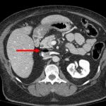
This photo shows the inferior vena cava of a patient with bilateral IO line insertions (black bubble at the top of the round IVC). During transport, one line was inadvertently disconnected and probably entrained some air. There was no adverse clinical effect, but if the problem is not recognized and the line closed, there could be.

Complications of Splenic Embolization for Trauma

Angioembolization has become a common procedure that can increase the likelihood of success for nonoperative management for splenic trauma. It does have its own set of complications to be aware of, however.

The most obvious complication is mechanical injury to the femoral artery. This occurs in 1 to 3% of patients. It is more common in the very young (small caliber artery) and the elderly (arteries of stone). Rarely, the substance or device that is used for the embolization may migrate or end up on the wrong spot, infarcting something important.

A common issue that occurs is infarction of portions of the spleen. This is actually the desired effect, as it stops the bleeding. Most of the time, we are unaware of the changes that take place in the spleen post-procedure. But every once in a while we get a repeat CT scan days or weeks down the road and see some very interesting things.

The most common finding is a splenic infarct alone. This is an area of the spleen, sometimes wedge shaped, that does not take up contrast. This is normal. In some cases, gas bubbles are seen within the spleen parenchyma, usually within the infarcted area. In others, large areas of gas are present, and an air-fluid level may also be seen. This is definitely not normal.

Tiny bubbles are normal after this procedure, and can be ignored if the patient does not appear ill and does not have any systemic evidence of inflammation or sepsis. On the other hand, big bubbles or air-fluid levels probably indicate a developing splenic abscess, and the patient will usually appear ill and have a high WBC count. Unfortunately, the only treatment for this is splenectomy. Insertion of drainage catheters does not work and the patient will only become sicker if it is attempted.

Top 10 Worst Complications: #1 Nasocerebral Tube

Minor complications from nasogastric tube insertion occur relatively frequently. Emesis is fairly common when the gag reflex is stimulated by the tube in the back of the oropharynx. An infrequent but possibly fatal one is insertion through the cribriform plate. 

The cribriform plate is located directly posterior to the nares and is part of the ethmoid bone. It is very porous in nature and weaker than the surrounding portions of the ethmoid. It is easily fractured, and can be seen is association with basilar skull fractures. This is one source for rhinorrhea in patients with these fractures.

Cribriform fracture is a contraindication to unprotected insertion of a nasogastric tube. If you look at the sagittal section below, the plate lies directly behind the nares. When inserting the NG tube, we are usually taught to aim the tube straight back. Unfortunately, this aims it directly at the cribriform. If a fracture is present, it is possible that you may be inserting a nasocerebral tube!

Cribriform plate - sagittal section

The usual symptoms when this occurs consist of immediate neurologic deterioration to coma, and a unilateral or bilateral blown pupil. The tube must not be withdrawn, because it will cause significant injury to the base of the brain. A stat neurosurgical consultation must be obtained, and if the patient is salvageable, the tube must be withdrawn through a craniectomy.

To avoid this dreaded complication, identify patients at risk for cribriform injury. They are:

  • patients with signs of trauma from eyebrows to zygoma
  • comatose patients
  • patients with signs of basilar skull fracture (Battle’s sign, raccoon eyes, oto- or rhinorrhea)

If your patient is at risk, follow these guidelines:

  • first, does the patient really need a gastric tube?
  • if comatose, insert an orogastric tube
  • if awake, don’t put the tube in their mouth, as they will gag continuously. Instead, place a lubricated, curved nasal airway. Then lube up a slightly smaller Salem sump tube and pass it through the airway.

The VIP (Very Important Person) Syndrome in Healthcare

The VIP syndrome occurs in healthcare when a celebrity or other well-connected “important” person receives a level of care that the average person does not. This situation was first documented in a paper published in the 1960s which noted that VIP patients have worse outcomes.

VIPs have the expectation that they can get special access to care and that the care will be of higher quality than that provided to others. Healthcare providers often grant this extra access, in the form of returned phone calls and preferential access to their clinic or office. The provider tries to provide a higher quality of care by ordering additional tests and involving more consultants. This idea ignores the fact that we already provide the best care we know how, and money or fame can’t buy any better.

Unfortunately, trying to provide better care sets up the VIP for a higher complication rate and a greater chance of death. Healthcare consists of a number of intertwined systems that, in general, have found their most efficient processes and lowest complication rates. Any disturbance in this equilibrium of tests, consultants, or nursing care moves this equilibrium away from its safety point.

Every test has its own set of possible complications. Each consultant feels compelled to add something to the evaluation, which usually means even more tests, and more possible complications. And once too many consultants are involved, there is no “captain of the ship” and care can become fragmented and even more inefficient and dangerous.

How do we avoid the VIP Syndrome? First, explain these facts to the VIP, making sure to impress upon them that requesting or receiving care that is “different” may be dangerous to their health. Explain the same things to all providers who will be involved in their care. Finally, do not stray from the way you “normally” do things. Order the same tests you usually would, use the same consultants, and take control of all of their recommendations, trying to do things in your usual way. This will provide the VIP with the best care possible, which is actually the same as what everybody else gets.

Reference: “The VIP Syndrome”: A Clinical Study in Hospital Psychiatry. Weintraub, Journal of Mental and Nervious Disease, 138(2): 181-193, 1964.