Tag Archives: bucket handle

Updated: How To Detect Bucket Handle Injuries With CT

A bucket-handle injury is a relatively uncommon complication of blunt trauma to the abdomen. It only occurs in a few percent of patients, but is much more likely if they have a seat belt sign.  The basic pathology is that the bowel mesentery (small bowel of sigmoid colon) gets pulled away from the intestinal wall.

This injury is problematic because it may take a few days for the bowel itself to die and perforate. Patients with no other injuries could potentially be discharged from the hospital before they become overtly symptomatic, leading to delayed treatment.

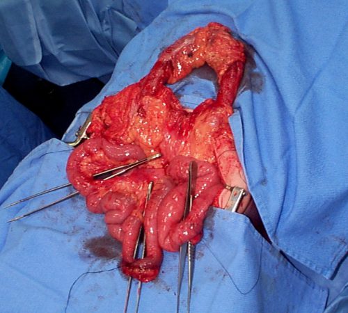
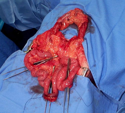
Here’s an image from my personal collection with not one, but four bucket-handle injuries.
Typical patients with suspected blunt intestinal injury are observed with good serial exams and a daily WBC count. If this begins to rise after 24 hours, there is a reasonable chance that this injury is present.

CT scan has not really been that reliable in past studies. There may be some “dirty mesentery”, which is contused and has a hematoma within it. But without a more convincing exam, it is difficult to convince yourself to operate immediately on these patients.

A paper was published by a group of radiologists at Duke University. It appears to be a case report disguised as a descriptive paper. It looks like they picked a few known bucket-handle injuries from their institution and back-correlated them with CT findings.

The authors called out the usual culprits:

  • Fluid between loops of bowel
  • Active bleeding in the mesentery
  • Bowel wall perfusion defects

But they also noted that traumatic abdominal wall hernias were highly associated with seat belt sign as well. These are rare, but should bring intestinal injury to mind when seen.

With newer scanners, radiologists are better able to detect subtle areas of hypoperfusion as well. This is a fairly good indicator of injury, especially when adjacent bowel appears normally perfused. Here are two examples. The black arrows denote active extravasation, and the white ones an area of hypoperfusion.
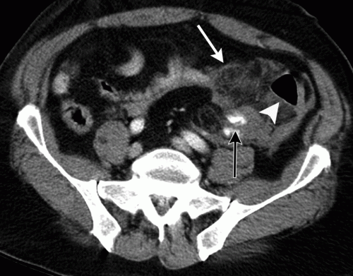
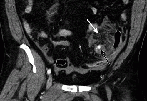
The authors add bowel wall hypoperfusion as another finding that may point to a bucket-handle type injury.

A recent paper demonstrates the value of the current generation of high-quality scanners. A collection of California and Denver centers implemented a multicenter, prospective, observational study of patients with seat belt signs. The developed a list of positive findings, which included:

  • abdominal wall soft tissue contusion (radiographic seat belt sign)
  • free peritoneal fluid
  • bowel wall thickening
  • mesenteric stranding
  • mesenteric hematoma
  • bowel dilation
  • pneumatosis
  • pneumoperitoneum

A total of 754 patients with visible seat belt sign were enrolled and all went to CT scan. Any of the findings listed above were associated with a statistically significant likelihood of hollow viscus injury. The highest likelihood was associated with:

  • free peritoneal fluid – 42x more likely
  • bowel dilation – 21x
  • free fluid with no solid organ injury – 20x
  • bowel wall thickening – 19x
  • radiographic seat belt sign – 3x

Any of the radiographic findings strongly suggested that an injury could be present. However, if none were present, it was very unlikely that there were any significant injuries. The authors suggested that if such patients had no other injuries requiring hospitalization, they could potentially be discharged home. However, those patients should be counseled to return for evaluation immediately if they have any change in their abdominal or systemic status.

Bottom line: Some patients with a visible seat belt sign might be eligible for discharge from the ED if they have a totally negative abdominal CT and no other injuries requiring hospitalization. If they have any finding, they should be admitted for observation.

If your patient has an unconcerning exam and any of the findings listed above, perform serial exams and get a WBC the next morning. If the exam worsens, operate. If the WBC rises, consider laparoscopy to see if you need to make a bigger incision. And if you see any evidence of hypoperfused bowel, consider laparoscopy right away. 

References:

  • Excluding Hollow Viscus Injury for Abdominal Seat Belt Sign Using Computed Tomography. JAMA Surg. 2022 Sep 1;157(9):771-778. doi: 10.1001/jamasurg.2022.2770. PMID: 35830194; PMCID: PMC9280606.
  • CT findings of traumatic bucket-handle mesenteric injuries. Am J Radiol 209:W360-@364, 2017.
  • Multidetector CT of blunt abdominal trauma. Radiology 265(3):678–693, 2012.

What Is: Bucket Handle Injury Of The Intestine

A bucket handle injury is a type of mesenteric injury of the intestine. The intestine itself separates from the mesentery, leaving a devascularized segment of bowel that looks like the handle on a bucket (get it?).

These injuries can occur after blunt trauma to the abdomen. The force required is rather extreme, so the usual mechanism is motor vehicle crash. In theory, it could occur after a fall from a significant height, and I have seen one case from a wood fragment that was hurled at the abdomen by a malfunctioning lathe.

The mechanics of this injury are related to fixed vs mobile structures in the abdomen. Injuries tend to occur adjacent to areas of the intestine that are fixed, such as the cecum, ligament of Treitz, colonic flexures and rectum. During sudden deceleration, portions of the intestine adjacent to these areas continue to move, pulling on the nearby attachments. This causes the intestine itself to pull off of its mesentery.

Source: personal archive. Not treated at Regions Hospital

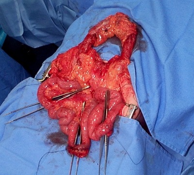
The picture above shows multiple bucket handle injuries in one patient. There are 3 injuries in the small bowel, and one involving the entire transverse colon. Note the obviously devascularized segment at the bottom center of the photo.

The terminal ileum is the most common site for bucket handle tears. Proximal jejunum, transverse colon, and sigmoid colon are other possible areas. In my experience, the driver is more likely to sustain an injury to the terminal ileum, and the front passenger to the sigmoid. I think this is due to the location of the buckle, but it’s just my own observation.

Always think about the possibility of this injury in patients with very high speed decelerationSeat belt marks have a particularly high association with this injury. If your patient has an abnormal exam in the right lower quadrant, or if the CT shows unusual changes there (“dirty” mesenteric fat, thickened bowel wall, extravasation), I recommend a trip to the OR. In these cases, an injury will nearly always be present. And since the affected intestine may take a few days to die and leak, look for an unexplained rise in WBC beginning on hospital day 2.

Related posts:

Bucket Handle Injuries Of The Intestine

Bucket Handle Injury

A bucket handle injury is a type of mesenteric injury of the intestine. The intestine itself separates from the mesentery, leaving a devascularized segment of bowel that looks like the handle on a bucket (get it?).

These injuries can occur after blunt trauma to the abdomen. The force required is rather extreme, so the usual mechanism is motor vehicle crash. In theory, it could occur after a fall from a significant height, and I have seen once case where a wood fragment was hurled at the abdomen by a malfunctioning lathe.

The mechanics of this injury are related to fixed vs mobile structures in the abdomen. Injuries tend to occur adjacent to areas of the intestine that are fixed, such as the cecum, ligament of Treitz, colonic flexures and rectum. During sudden deceleration, portions of the intestine adjacent to these areas continue to move, pulling on the nearby attachments. This causes the intestine itself to pull off of its mesentery.

The terminal ileum is the most common site for bucket handle tears. Proximal jejunum, transverse colon, and sigmoid colon are other possible areas. The picture above shows multiple bucket handle injuries in one patient. There are 3 injuries in the small bowel, and one involving the entire transverse colon. Note the obviously devascularized segment at the bottom center of the photo.

Always think about the possibility of this injury in patients with very high speed decelerationsSeat belt marks have a particularly high association with this injury. If your patient has an abnormal exam in the right lower quadrant, or if the CT shows unusual changes there (“dirty” mesenteric fat, thickened bowel wall, extravasation), I recommend a trip to the OR. In these cases, an injury will nearly always be present.

Related posts:

Source: personal archive. Not treated at Regions Hospital

Bucket Handle Injury – Part 2

Yesterday, I wrote about the basics of bucket handle injuries of the intestine. Today, I’ll deal with diagnosing them.

An understanding of the mechanism of injury and a good physical exam are paramount. If the patient took a significant blow to the abdomen, especially in a motor vehicle crash (lap belt), be very suspicious. Any abdominal pain is of concern, particularly in the right lower quadrant (most common injury location). If a CT is indicated and there are focal changes in the mesenery or bowel wall, a trip to the OR is advised.

In some patients, the bowel is devascularized and takes 2-3 days to become necrotic. They experience slowly increasing focal pain, and once this develops it’s time to go to the operating room.

Intubated and/or comatose patients can be problematic in making this diagnosis. There is no physical exam, so the trauma professional has to rely on surrogates. The white blood cell (WBC) count is very helpful. The WBC count is typically elevated into the 15,000-20,000 range immediately after trauma, and declines to normal within about 12 hours. If it begins to climb again after 24 hours, especially if it exceeds 20,000, an intestinal injury is likely.

CT scan and abdominal ultrasound are also helpful. A repeat CT scan may show a change in the volume of fluid, or a change in its character. If the amount of fluid increases significantly, or if a fluid bi-layer is seen, a bucket handle injury is very likely. These findings are pertinent in awake patients as well, but the physical exam usually makes use of these diagnostics unnecessary.

Related posts:

Bucket Handle Injury – Part 1

Bucket Handle Injury

A bucket handle injury is a type of mesenteric injury of the intestine. The intestine itself separates from the mesentery, leaving a devascularized segment of bowel that looks like the handle on a bucket (get it?).

These injuries can occur after blunt trauma to the abdomen. The force required is rather extreme, so the usual mechanism is motor vehicle crash. In theory, it could occur after a fall from a significant height, and I have seen once case where a wood fragment was hurled at the abdomen by a malfunctioning lathe.

The mechanics of this injury are related to fixed vs mobile structures in the abdomen. Injuries tend to occur adjacent to areas of the intestine that are fixed, such as the cecum, ligament of Treitz, colonic flexures and rectum. During sudden deceleration, portions of the intestine adjacent to these areas continue to move, pulling on the nearby attachments. This causes the intestine itself to pull off of its mesentery.

The terminal ileum is the most common site for bucket handle tears. Proximal jejunum, transverse colon, and sigmoid colon are other possible areas. The picture above shows multiple bucket handle injuries in one patient. There are 3 injuries in the small bowel, and one involving the entire transverse colon. Note the obviously devascularized segment at the bottom center of the photo.

Always think about the possibility of this injury in patients with very high speed decelerations. Seat belt marks have a particularly high association with this injury. If your patient has an abnormal exam in the right lower quadrant, or if the CT shows unusual changes there (“dirty” mesenteric fat, thickened bowel wall, extravasation), I recommend a trip to the OR. In these cases, an injury will nearly always be present.

Tomorrow: These injuries can be subtle in an awake patient with a reliable exam. On Friday I’ll write about how you can detect it in unconscious patients.

Source: personal archive. Not treated at Regions Hospital