Tag Archives: Blunt abdominal trauma

How To Detect Bucket-handle Intestinal Injuries With CT Scan

A bucket-handle injury is a relatively uncommon complication of blunt trauma to the abdomen. It only occurs in a few percent of patients, but is much more likely if they have a seat belt sign.  The basic pathology is that the bowel mesentery (small bowel of sigmoid colon) gets pulled away from the intestinal wall.

This injury is problematic because it may take a few days for the bowel itself to die and perforate. Patients with no other injuries could potentially be discharged from the hospital before they become overtly symptomatic, leading to delayed treatment.

Here’s an image from my personal collection with not one, but four bucket-handle injuries.
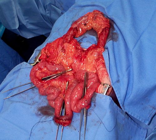
Typical patients with suspected blunt intestinal injury are observed with good serial exams and a daily WBC count. If this begins to rise after 24 hours, there is a reasonable chance that this injury is present.

CT scan has not really been that reliable in past studies. There may be some “dirty mesentery”, which is contused and has a hematoma within it. But without a more convincing exam, it is difficult to convince yourself to operate immediately on these patients.

A paper was published by a group of radiologists at Duke University. It appears to be a case report disguised as a descriptive paper. It looks like they picked a few known bucket-handle injuries from their institution and back-correlated them with CT findings.

The authors called out the usual culprits:

  • Fluid between loops of bowel
  • Active bleeding in the mesentery
  • Bowel wall perfusion defects

But they also noted that traumatic abdominal wall hernias were highly with the injury as well. These are rare, but should bring intestinal injury to mind when seen.

With newer scanners, radiologists are better able to detect subtle areas of hypoperfusion as well. This is a fairly good indicator of injury, especially when adjacent bowel appears normally perfused. Here are two examples. The black arrows denote active extravasation, and the white ones an area of hypoperfusion.
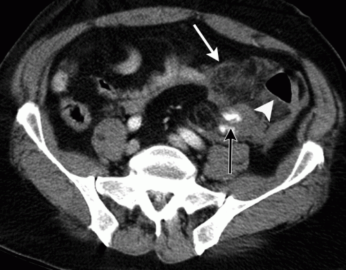
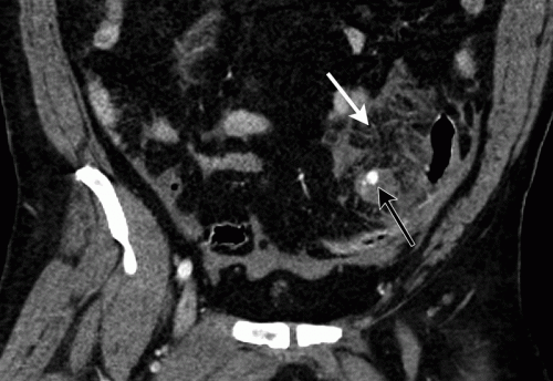
The authors add bowel wall hypoperfusion as another finding that may point to a bucket-handle type injury

Bottom line: Hold the phone! Don’t change your practice yet. This paper is not able to demonstrate how good this radiographic sign is. Looking at other radiology literature, the specificity is about 90%. But remember, that means that if they don’t have the CT finding, that’s true only 90% of the time.

Unfortunately the sensitivity is only 10%. So if you see it on the scan, they’ve got a 1 in 10 chance of actually having the injury. That’s not good enough for me to run to the operating room.

Here’s what I recommend: if your patient has an unconcerning exam and any of the usual culprits (pelvic fluid, inter-loop fluid, dirty mesentery, thickened bowel loops, abdominal wall hernia), perform serial exams and get a WBC the next morning. If the exam worsens, operate. If the WBC rises, consider laparoscopy to see if you need to make a bigger incision. And if you see this new kid on the block, the hypoperfused bowel, consider laparoscopy right away. 

I’m sure the radiologists and the technology will keep getting better. But for now, blunt intestinal injury still requires patience, perceptiveness, and a little luck.

References:

  • CT findings of traumatic bucket-handle mesenteric injuries. Am J Radiol 209:W360-@364, 2017.
  • Multidetector CT of blunt abdominal trauma. Radiology 265(3):678–693, 2012.

EAST 2018 #5: Predicting Absence Of Pediatric Abdominal Injury

More on prediction systems today! The authors of this abstract used good old mathematics, albeit very fancy math, instead of a machine learning algorithm. The specifics of this tool were described in an article published in JACS earlier this year (see reference).

The authors were interested in finding a way to decrease the use of CT scan for evaluating blunt abdominal trauma in children. After developing the model using prospectively collected data from 14 Level I pediatric trauma centers, they sought to validate it using a public dataset from the Pediatric Emergency Care Applied Research Network (PECARN). This dataset contained more than 2,400 records, and included 10% of patients who had an intra-abdominal injury (IAI), and 2.5% with an IAI that required intervention (IAI-I).

Here are the factoids:

  • There were five prediction rule variables: complaint of abdominal pain, tenderness / distension / or contusion on exam, abnormal chest x-ray, AST > 200, elevated pancreatic enzymes)
  • Prediction rule sensitivity was 98% and specificity was 37% for IAI, and 100% / 35% for IAI-I
  • The negative predictive value for finding any abdominal injury was 99.3%, and for injury requiring intervention was 100%
  • Unfortunately, nearly half of the very low risk children underwent CT scanning anyway!

Bottom line: This is a nice validation study for a well-designed prediction tool. It builds on previous work published earlier this year. The variables make clinical sense. Although the number of patients with injury were relatively small, I believe these results should be considered for incorporation in our blunt pediatric trauma evaluation protocols now!

Here are some questions for the authors to consider before their presentation:

  • The liver function and pancreatic enzyme tests results take some time to perform. How much do they contribute to the negative predictive value, since they are relatively uncommon injuries?
  • What are considered abnormal chest x-ray findings?
  • How do you recommend incorporating this into the care of trauma activation patients? Wait for 30 minutes in the trauma bay for the lab tests to come back? Evaluation in patients undergoing a more routine evaluation for abdominal trauma would not be unduly delayed.
  • Be prepared to explain how you derived the decision rule in very simple language.

References:

  • EAST 2018 Podium paper #7.
  • Identifying Children at Very Low Risk for Blunt Intra-Abdominal Injury in Whom CT of the Abdomen Can Be Avoided Safely. JACS 224(4):449-458.

“Found Down”: Do We Need To Worry About The Abdomen?

It’s that dreaded mechanism of injury: “found down.” What really happened? Did they fall, or get assaulted? Or did the patient suffer a medical problem that led to them falling down? Trauma professionals rely heavily on what I call “context.” Is the patient elderly and frail? Are they intoxicated? What was their location when found? Are there pre-existing medical conditions?

All of these factors allow us to begin building a story in our mind that tells us what might have happened, and what the injuries might be. But with little or no context, we are flying by the seat of our pants. We end up suspecting everything, which means we image everything. With CT scans. And IV contrast. There is always a chance that we can add to the harm already suffered by this patient, or waste time and money. 

The abdomen is a black box in a patient with an unreliable or absent physical exam. The emergency medicine group at Cedars-Sinai in Los Angeles looked at the utlity of CT scanning the abdomen in this group of patients. They retrospectively reviewed 10 years of their data. They found 342 patients, of which 154 underwent some type of abdominal imaging (CT, FAST).

Here are the factoids:

  • About 60% had alcohol present, and 98% of those had a level greater than 0.08 g/dL
  • Overall mortality was 10%. 24 were trauma related (severe TBI, traumatic arrest in ED), and 9 were medical (CVA, sepsis)
  • 55% did not undergo any abdominal imaging, and their mortality was 6% (TBI, stroke, MI). None manifested a late abdominal injury.
  • Of the 45% who did have abdominal imaging, 57% had CT, 24% FAST, and 19% both
  • 14 patients in the imaged group had a positive abdominal CT, but all were minor (Abbreviated Injury Score (AIS) <3)
  • 5 patients had a positive FAST, and all had an abdominal AIS<3

Bottom line: Patients who are “found down” seldom have significant intra-abdominal injuries. Keep in mind that this is a small study group, but it does seem to correlate with personal experience and reviews of many charts. Although you can’t completely ignore the abdomen in this group of patients, you should place a higher priority on head and neck trauma, or CVA/sepsis in patient without other obvious injury.

Related posts:

Reference: Abdominal injuries in the “found down”: is imaging indicated? J Am Col Surg 221(1):17-25, 2015.

Initial Evaluation of Blunt Abdominal Trauma – Update

This preliminary EAST Practice Management Guideline was presented and discussed at the 23rd Annual Scientific Assembly of the Eastern Association for the Surgery of Trauma.

The EAST practice guideline regarding evaluation of blunt abdominal trauma was first published in 2001. It was updated by performing a new literature search spanning 1998 to 2009. A total of 33 new articles were reviewed to provide material for the revised guideline. As usual, the number of high quality references (3 Class I and 11 Class II) were outnumbered by lower quality Class III references (19).

For information on classes of data and levels of recommendations, please refer to the Primer on Evidenced Based Medicine on the EAST website.

Important: These guidelines are preliminary and may undergo further minor revision, so the final version may be slightly different than described here!

The Level I recommendations remained basically the same, with one modification (bolded below):

  1. FAST may be considered as the initial diagnostic modality to exclude hemoperitoneum.
  2. Exploratory laparotomy is indicated in hemodynamically unstable patients with a positive FAST. In hemodynamically stable patients with a positive FAST, follow-up CT scan permits nonoperative management of select injuries.
  3. Exploratory laparotomy is indicated for patients with a positive DPL and hemodynamic instability.

There was some interesting discussion about the continued utility of DPL. Some audience members felt that this was an outdated technique. Others pointed out that not all surgeons work in a Level I or II trauma center, and that FAST may not be available to them, so the technique remains relevant. Additionally, these guidelines may be used abroad where more advanced diagnostic testing is not as readily available, so it was recommended that the DPL language be retained.

The Level II recommendations are:

  1. When DPL is used, clinical decisions should be made on the basis of the presence of gross blood on initial aspiration (i.e. 10ml) or microscopic analysis of lavage effluent.
  2. Surveillance studies (i.e. DPL, CT scan, repeat FAST) should be considered in hemodynamically stable patients with indeterminate FAST results.
  3. CT scanning is recommended for the evaluation of hemodynamically stable patients with equivocal findings on physical examination, associated with neurologic injury, or multiple extra-abdominal injuries. Under these circumstances, patients with a negative CT should be admitted for observation.
  4. CT scanning is the diagnostic modality of choice for nonoperative management of solid visceral injuries.
  5. In hemodynamically stable patients, DPL and CT scanning are complementary diagnostic modalities.
  6. Contrast enhanced ultrasound (CEUS) is more sensitive than non-contrast ultrasound in the detection of solid organ injury. Many members of the audience were not familiar with this technique. I will comment on it in a later blog entry.
  7. In the patient at high risk for intra-abdominal injury (e.g. multiple orthopedic injuries, severe chest wall trauma, neurologic impairment) a CT scan should be considered in hemodynamically stable patients, even after a negative FAST.

Finally, the Level III recommendations are:

  1. Objective testing (i.e. FAST, DPL, CT scanning) is indicated for patients with abnormal mentation, equivocal findings on physical examination, multiple injuries, concomitant chest injury, or hematuria.
  2. Patients with seat belt sign should be admitted for observation and serial physical examination. The presence of intraperitoneal fluid on FAST or CT scan in a patient with seat belt sign suggests the presence of an intra-abdominal injury that may require surgery.
  3. CT scanning is indicated for suspected renal injuries.
  4. In hemodynamically stable patients with a positive DPL, a CT scan should be considered, especially in the presence of pelvic fracture or suspected injuries to the genitourinary tract, diaphragm or pancreas.
  5. Patients with free fluid and no solid organ injury on CT should be considered for laparotomy. Alternatively, laparoscopy or DPL may aid in diagnosis of bowel injury. Patients with no head injury and clear mentation may be followed by serial exams.