Tag Archives: aorta

Early Vs Delayed Thoracic Endovascular Repair

Back in the day, the only way to fix a broken thoracic aorta was via left thoracotomy.  This was a big procedure, with the possibility of several major complications, with postop paraplegia being one of them. At the time, there was a debate about whether the procedure should be done immediately versus waiting until the patient was well-resuscitated. The concern was that death was nearly certain if the aortic lesion progressed.

We learned that temporizing with strict blood pressure control worked wonders at protecting the patient. Although many of these injuries were managed within hours, a growing number were delayed by a few days to improve outcomes.

Nowadays, thoracic endovascular aortic repair (TEVAR) is routine and much less morbid than the open procedure. However, the same question arises: do it early or wait a while? Interestingly, not one but two analyses have been published on this very topic in the last four months!

The first is from an international research group that searched the usual databases and initially found 921 records. They included only clinical trials or cohort studies with ten or more adult patients that could be stratified as early (within 24 hours) or late (after 24 hours) intervention. After applying these criteria, only seven studies remained for analysis.

There were 3,757 patients with early repairs, compared to 1,238 undergoing late repair. The presenting demographics and injury grades were similar in each group. However, the short-term mortality was significantly higher (1.9x) in the early TEVAR group. Additionally, ICU length of stay was significantly longer (3 days) in the late TEVAR group.

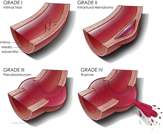
The second paper was presented as a quick-shot at last year’s AAST meeting. It is from a group of researchers from our big Boston trauma centers and the Netherlands. They used four years of data from the TQIP database, giving them extra information unavailable in the first study. They specifically looked at patients with grade II or III injuries. Here is the grading scale:
Here are the factoids:

  • A total of 1,339 patients were studied, with about three-quarters in the early TEVAR group
  • Median time to TEVAR was 4 hours in the early group and 65 hours in the late group
  • Patients in the early group were significantly less likely to have brain or liver injuries
  • ISS was similar in both groups
  • The early TEVAR group had significantly higher in-hospital mortality (16% vs. 5%), significantly higher risk of ARDS (7.6% vs. 2.1%), but significantly shorter ICU stay (7 vs 10 days)
  • When patients who died within the first 24 hours were excluded, the in-hospital mortality remained significantly higher, and the ICU and hospital lengths of stay were significantly shorter

Bottom line: Some society guidelines began recommending delayed TEVAR in 2015. This study did not detect any trend toward this, however. Using different methods and databases, these two studies identified nearly identical mortality and ICU trends in large groups of patients. The mortality trends do not appear to be related to injury grade, overall injury severity, or the presence of head injury. 

Taken together, this suggests that we need to rethink the timing of TEVAR in patients with grade II or III injuries. The best timing still needs to be defined, but it appears to be beyond 24 hours. Centers performing this procedure should review their results and consider extending procedure timing as additional research is done to define the ideal time interval.

References:

  1. Early Versus Delayed Thoracic Endovascular Aortic Repair for Blunt Traumatic Aortic Injury: A Systematic Review and Meta-Analysis. Cureus. 2023 Jun 28;15(6):e41078. doi: 10.7759/cureus.41078. PMID: 37519486; PMCID: PMC10375940.
  2. Early Versus Delayed Thoracic Endovascular Aortic Repair for
    Blunt Thoracic Aortic Injury: A Propensity Score-Matched Analysis. Ann Surg 278:e848-e854, 2023.


What Is A Wide Mediastinum Anyway?

Trauma professionals are always on the lookout for injuries that can kill you. Thoracic aortic injury from blunt trauma is one of those injuries. Thankfully, it is uncommon, but it can certainly be deadly.

One of the screening tests used to detect aortic injury is the old-fashioned chest xray. This test is said to be about 50% sensitive, with a negative predictive value of about 80%. However, the sensitivity is probably decreasing and the negative predictive value increasing due to the rapidly increasing number of obese patients that we see.

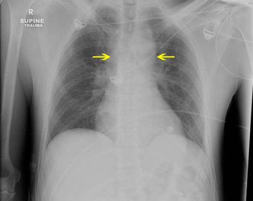
A wide mediastinum is defined as being > 8cm in width. In this day and age of digital imaging, you will need to use the measurement tool on your workstation to figure this out.

Unfortunately, it seems like most chest xrays show wide mediastinum these days. What are the most common causes for this?

  • Technique. The standard xray technique used to reduce magnification of the anterior mediastinum (where the aortic arch lives) is a tube distance of 72 inches from the patient, shot back to front. We can’t do this for trauma patients because we can’t stand them up and are reluctant to prone them. The standard trauma room technique is 36 inches from the patient shot front to back. This serves to magnify the mediastinal image and make it look wide.
  • Obesity. The more fat in the mediastinum, the wider it looks. The more fat on the back, the further the mediastinum is from the xray plate and the greater the magnification.
  • Other mediastinal blood. Major blunt trauma to the chest can cause bleeding from small veins in the mediastinum, making it look wide.
  • Thymus. Only in kids, though.
  • Aortic injury. Last but not least. Only a few percent of people with wide mediastinum will actually have the injury.

If you encounter a wide mediastinum on chest xray in a patient with a significant mechanism for aortic injury, then they should be screened using helical CT.

AAST 2019 #8: Timing Of Thoracic Aortic Injury Repair

Over the past two decades, there has been a massive swing from open repair of blunt thoracic aortic injury to thoracic endovascular aortic repair (TEVAR). Although technically a bit more complex, it has decreased both morbidity and mortality significantly. The usual push in fresh trauma patients is to take care of all the life-threatening injuries as soon as possible. And from the days of the open thoracic procedure, this was generally warranted.

However, the optimal timing of repair during the age of TEVAR is not as clear. Is it really necessary to go crashing into the angio or hybrid suite to get this taken care of? Or should it wait until the patient is not as physiologically damaged? The group at University of Texas at San Antonio looked at experience in the National Trauma Databank for some guidance. They reviewed four years of data from 2012 to 2015. Patients who arrested in or prior to arrival in the ED were excluded. Mortality was the primary outcome of interest, but complications and hospital length of stay (LOS) was also noted.

Here are the factoids:

  • Nearly 6,000 patients with blunt thoracic aortic injury were identifed, and 1,930 (33%) underwent TEVAR, 2% were opened, and 65% were managed nonoperatively
  • Looking only at TEVAR patients, 69% underwent the procedure within 24 hours, 24% after 24 hours, and the remainder were not recorded (!)
  • Mortality was significantly higher in the early TEVAR group (6.4% vs 2.1%)
  • Hospital LOS was significantly shorter in the early TEVAR group (18 vs 22 days)
  • Logistic regression controlling for hypotension, severe TBI, ISS and older age confirmed the significantly lower mortality in the delayed group

The authors concluded that delayed (>24 hrs) TEVAR was associated with decreased mortality but longer length of stay.

This is a nice, clean abstract to read. The hypothesis and results are easy to understand and make sense. And it’s exactly the kind of poster that makes you think a bit. 

The only real downside is that it is an NTDB study, so there is very limited ability to go back and tease out why these results should be true. These results should push the authors to set up a more prospective study so they can figure out why this should be true. We can certainly speculate that it helps to temporize with good blood pressure control while cleaning up other major injuries and correcting deranged physiology. But one never knows until the right study is actually done.

Here are my questions for the presenter and authors:

  • Were you able to glean any insights into the associations you identified from the other data in the NTDB records you used? This could help design a really good study to see if your impressions are true.
  • The fact that a quarter of patients had TEVAR at an unknown time throws a big monkey wrench in your results. Can you use any statistical tricks to see if assuming they were either early or late would influence your results. Is it possible that this unknown group could completely neutralized your study?

I’m very excited by this one, and I don’t normally get too excited by posters. Great work!

Reference: Timing of repair of blunt traumatic thoracic aortic injury: results from the National Trauma Databank. AAST 2019, Poster #5.

Long-Term Experience With Endovascular Aortic Repair For Trauma

For decades, the treatment of blunt injury to the thoracic aorta was open repair. The big debate at the time was use of cardiac bypass vs fast clamp and sew. But starting in 1997 with the introduction of thoracic endovascular aortic repair (TEVAR) of this injury, we have rapidly moved to the point where most traumatic aortic injuries are repaired using this technique.

A report that was written nearly a decade ago indicated a relatively high complication rate for the procedure. Graft complications were reported in 18% of patients, with 14% showing endoleaks. Stroke and left arm ischemia were also reported.

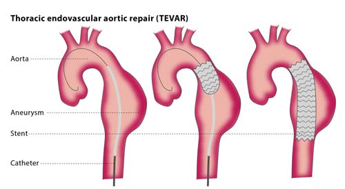
The diagram above shows insertion for management of an aneurysm, but the technique is similar for trauma. Blunt aortic injury occurs closer to the left subclavian artery and care must be taken to place the endograft closer to but not covering its orifice.

As the insertion systems and stents improved, short term events have been on the decline. Unfortunately, long term followup data has been hard to come by.

Until now. An article that is not yet in print reports 11 years of experience and followup with patient undergoing TEVAR at the ShockTrauma center in Baltimore.

Here are the factoids:

  • 88 patients underwent TEVAR during the study period, all from blunt trauma
  • Average ISS was 38, showing these patients were severely injured
  • Overall mortality was 7%, but none was due to the TEVAR procedure
  • TEVAR-related complication rate was 9% Endoleaks at the ends of the graft occurred in 4 patients, and all required repair. There were 4 other minor leaks that resolved on their own.
  • 26 had all or part of the left subclavian orifice covered at initial operation. None developed ischemia, although 2 had a prophylactic carotid-subclavian bypass before TEVAR.
  • The longest followup imaging occurred 8 years after the procedure. No long-term complications were noted.

Bottom line: TEVAR has essentially replaced open repair of the aorta, except in special cases. We continue to learn from our experience, and the complication rate is still falling. Other than endoleaks recognized in the postop period, most other complications rarely occur. Long term followup is poor, but in the patients who do return, there were no complications. But remember, this is an expected sampling bias. If the patient had major problems and/or died, they would just be lost to followup. We would never know.

EAST Guidelines: Blunt Traumatic Aortic Injury

The Eastern Association for the Surgery of Trauma (EAST) has been helping trauma professionals through the publication of practice guidelines for more than 15 years. Members of EAST donate their time to review reams of literature, good and bad, to try to determine the answers to common or puzzling clinical questions.

Why follow a practice guideline? Quite simply, if properly developed, a guideline represents our best understanding of the “correct” answer to the question posed. And as many of you who follow this blog already know, things that “seem to make sense” frequently are totally wrong. Your own experience is poignant, but the pooled experience of the many others who contributed to research on the topic in question is much more significant.

So on the the practice guideline for blunt traumatic aortic injury (BTAI). This one answers three questions. I will list each, followed by the conclusions reached through the literature review.

1. In patients with suspected BTAI, which diagnostic modality should be chosen: CT angiography of the chest, or conventional catheter angiography?

  – Catheter angiography was the standard for decades. When the first EAST guideline on this topic was released 15 years ago, CT angiography was only a level III recommendation because experience with it was lacking. CT technology has advanced rapidly, with multiple detectors, helical scanning, and incredible computing power. Although the quality of the evidence is somewhat low, the panel strongly recommends the use of CT angiography due to its ready availability, speed, low invasiveness, and ability to detect and define other injuries.

2. Should endovascular or open repair be selected in order to minimize stroke, renal failure, paraplegia, and death?

  – Once again, the quality of available data is so so. However, it was possible to detect differences in outcome in comparative studies. The panel strongly recommends the use of endovascular repair in patients who do not have contraindications due to its lower blood loss, mortality, and paraplegia, and equivalent risk of stroke. Furthermore, it is performed more frequently now than open repair, and experience is thus greater at many institutions, further reducing complications.

3. Should the repair be performed immediately or delayed in order to minimize stroke, renal failure, paraplegia, and death?

  – Literature review revealed that the incidence of renal failure and paraplegia were lower with delayed repair, while renal failure was the same in patients with significant associated injuries. There was benefit to delaying repair until resuscitation was achieved and any other life threatening injuries were addressed. The panel recommends that delayed repair be carried out once these other conditions have been corrected. The procedure should not be delayed until the next morning for the convenience of the surgeons.

Related posts:

Reference: Evaluation and management of blunt traumatic aortic injury: a practice guideline from the Eastern Association for the Surgery of Trauma. 78(1):136-146, 2015.