Category Archives: Tips

Trauma Surgery Tip: How To See The Unseeable – The Answer

Yesterday I posed a scenario where the surgeon needed to see an area of an open abdomen (trauma laparotomy) that could not easily be visualized. Specifically, there was a question as to whether the diaphragm had been violated just anterior to the liver, just under the costal margin.

Short of putting your head in the wound, how can you visualize this area? Or some other hard to reach spot? Well, you could have an assistant insert a retractor and pull like crazy. However, the rib cage might not bend very well, and in elderly patients it may break. Not a good idea.

Some readers suggested breaking out the laparoscopy equipment and using the camera and optics to visualize. This is a reasonable idea, but expensive. Shouldn’t there be some good (and cheap) way to do this?

Of course, and there is. Think low tech. Very low tech. You just need to see around a corner, right. So get a mirror!

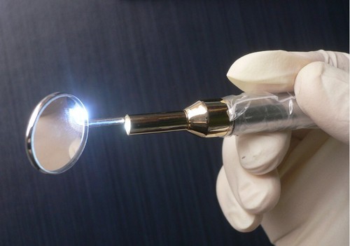
Every OR has some sterile dental mirrors lying around. Get one and have your assistant gently hold the liver down while you indirectly examine the diaphragm. Since you’re probably not a dentist, it may take a minute or two to get used to manipulating the mirror to see just what you want. But if you can manage laparoscopic surgery, you’ll get the hang of it quickly.

And if you need more light up in those nooks and crannies? Shine the OR light directly into the abdomen, then place a nice shiny malleable retractor into the area to reflect light into the area in questions. Voila!

Bottom line: A lot of the things that trauma professionals need to do in the heat of the moment will not be found in doctor, nurse, or paramedic books. Be creative. Look at the stuff around you and available to you. Figure out a way to make it work, and make $#!+ up if necessary.

Print Friendly, PDF & Email

Trauma Surgery Tip: How To See The Unseeable

Let me present a scenario and first see how you might solve this problem.

A young man presents with a gunshot to the abdomen in the right mid-back. He is hemodynamically stable, and you get a chest xray. It shows a small caliber slug in the right upper quadrant, but no hemo- or pneumothorax. He has peritoneal signs, so you whisk him off to the OR for a laparotomy.

As you prep the patient for the case, you can feel a small mass just above the right costal margin. You incise the area and produce a 22 caliber bullet. Of course, you follow the chain of evidence rules and pass it off for the police. As you explore the abdomen, it appears that there are no gross injuries. You are concerned, however, that there may be an injury to the diaphragm in proximity to the bullet.

So here’s the question: how can you visualize the diaphragm in this area? The bullet was located below the right nipple. But the diaphragm in this area is covered by the liver, and is parallel to the floor. You can’t seem to feel a hole with your fat finger. But short of putting your whole head in the wound, you just can’t get a good angle to see the area in question.

How would you do it? Please tweet or leave comments with your suggestions. I’ll provide the answer(s) tomorrow!

Print Friendly, PDF & Email

Stuff You Sterilize Other Stuff With May Not Be Sterile??

When one works in the trauma field, or medicine in general, we deal with the need for sterility all the time. We use equipment and devices that are sterile, and we administer drugs and fluids that are sterile. In surgery, we create sterile fields in which to use this sterile stuff.

In the past few years, we’ve come to the realization that the sterility we take for granted may not always be the case. There have been several cases of contaminated implanted hardware. And a few years ago, supposedly sterile injectable steroids were found to be contaminated with fungus, leading to several fatal cases of meningitis.

An article in the New England Journal of Medicine brings a bizarre problem to light: microbial stowaways in the topical products we use to sterilize things. Most drugs and infused fluids are prepared under sterile conditions. However, due to the antimicrobial activity of topical antiseptics, there is no requirement in the US that they be prepared in this way.

A number of cases of contamination have been reported over the years:

  • Iodophor – contamination with Burkholderia and Pseudomonas occurred during manufacture, leading to dialysis catheter infection and peritonitis
  • Chlorhexidine – contaminated with Serratia, Burkholderia and Ralstonia by end users, leading to wound infections, catheter infections, and death
  • Benzalkonium chloride – contaminated with Burkholderia and Mycobacteria by end users, causing septic arthritis and injection site infections

Bottom line: Nothing is sacred! This problem is scarier than you think, because our most basic assumptions about these products makes it nearly impossible for us to consider them when tracking down infection sources. Furthermore, they are so uncommon that they frequently may go undetected. The one telltale sign is the presence of infection from weird bacteria. If you encounter these bugs, consider this uncommon cause. Regulatory agencies need to get on this and mandate better manufacturing practices for topical antiseptics.

Reference: Microbial stowaways in topical antiseptic products. NEJM 367:2170-2173, Dec 6 2012.

Print Friendly, PDF & Email

Practical Tips: Transferring The Mangled Extremity

Managing the mangled extremity is both challenging and intense. There is always pressure to do all we can to save that threatened limb. But as you know, different levels of trauma centers have different capabilities and specialists that are needed to fully manage these injuries.

Level I centers have a comprehensive set of specialists to deal with the managed extremity, including trauma surgeons, vascular surgeons, orthopedic surgeons comfortable with complex injury, plastic surgeons, and interventional radiologists. The expectation is that a mangled extremity can be completely managed at such a center.

Level III centers have much more limited resources, and may only have a trauma surgeon to perform the initial evaluation. Definitive management can only occur after transfer to a Level I center.

Level II centers often find themselves in a kind of limbo. They have most of the specialties required, but those specialists may have varying comfort levels regarding addressing complex injuries. Some Level II centers may be able to keep these patients, but many will find that they need to transfer to their upstream Level I partner.

What do transferring trauma centers need to do before actually moving the patient? Here are some practical tips.

  • Evaluate quickly. The bottom line is to try to preserve function, so time is of the essence. Do a thorough evaluation of the anatomy, as well as vascular and neurologic status. These are the major determinants of salvageability.
  • Don’t ignore the rest of the patient. Make sure that injuries more critical than the extremity are identified and addressed. See the “Dang Factor!” below.
  • Make a decision. Now. Decide whether you need to transfer the patient based on your knowledge of your consultants’ skill levels and comfort.
  • Once you decide you will transfer, do no further imaging. It’s not going to change anything you do, and may not be very helpful to the receiving center.
  • Give IV antibiotics and the life-saving tetanus shot early.
  • Optimize salvageability. Do what you can to keep tissue healthy during the transfer. You must take transfer time into account for this! If you are sending your patient across town, just do it quickly. However, if he or she must travel long distance, there are a few more things to consider:
    • Try removing the tourniquet (if any). You’d be surprised at how many times the bleeding has stopped already. Or maybe wasn’t needed in the first place.
    • Selectively try to control bleeding if possible. Carefully ligate small vessels if you can. Don’t clamp and tie large masses of tissue.
    • Consider a vascular shunt. If there is an obvious large vessel injury, and if you have a trauma or vascular surgeon who is comfortable with inserting a vascular shunt, do it prior to transfer. This will increase the likelihood of salvage in long-distance transfers. But don’t waste a lot of time doing this! If you can’t get it done within about 30 minutes or so, don’t delay the transfer.
    • Quickly rinse off the area. Try to minimize the time that noxious stuff (dirt, gasoline, etc) is in contact with the tissues.
    • Splint well. You’ll need to be creative. But you don’t want additional tissue injury due to the extremity just flopping around.
  • Inquire about followup. Find out how the patient did, and discuss anything you could have done differently with the receiving center. As always, performance improvement is important!

Related posts:

Print Friendly, PDF & Email

Thoughts On Traumatic Hematuria: Part 1

I’ve seen a number of patients recently with bloody urine, and that is prompting me to provide some (written) clarity to others who need to manage this clinical problem. I’ll try to keep it organized!

There are two kinds of hematuria in trauma: blood that you can see with the naked eye, and…

Okay, so there’s only one. Trauma professionals do not care about microscopic hematuria. It does not change clinical management. Sure, your patient might have a renal contusion, but you won’t do anything about that. Or, he/she might have an infarcting kidney. And you can’t do anything about that. If you order a urinalysis, you might see a few RBCs. Don’t let this lead you down the path of looking for a source. You’ll end up ordering lots of tests and additional imaging, and generally will have nothing to show for it at the end. It’s not your job to spend good money on the very rare chance of finding something clinically significant.

Both of these specimens have blood in them. You can’t see it on the left, so don’t go looking for it with a microscope.

There are four sources of blood in the urine.

1. The first source does not generally cause hematuria, but can occasionally cause a few visible wisps of blood. That source is a urethral injury. The textbook teaching, and it’s good advice, is to look at the urethral meatus in your trauma patient, especially if you are contemplating insertion of a urinary catheter. If you see a few drops of blood, pause to consider. Sometimes, the blood is no longer visible, but might be present as a few well-placed drops on the patient’s underwear. So have a look at that, too, especially in patients with high risk injuries such as A-P compression pelvic fractures (think, lots of ramus fractures or pubic diastasis).

If you didn’t notice it and inserted the catheter anyway, you might see a few wisps of blood in the tubing as you place it. More often than not, this is just run of the mill irritation of the mucosa by the catheter, but always keep the possibility of an injury in mind.

Tomorrow, I’ll discuss the remaining three sources, and what to do about them.

Related posts:

Print Friendly, PDF & Email