This video is for nurses, phsyicians, medics, and anyone else who has to put one of these on. Looks simple, but there are some nuances you need to know about.
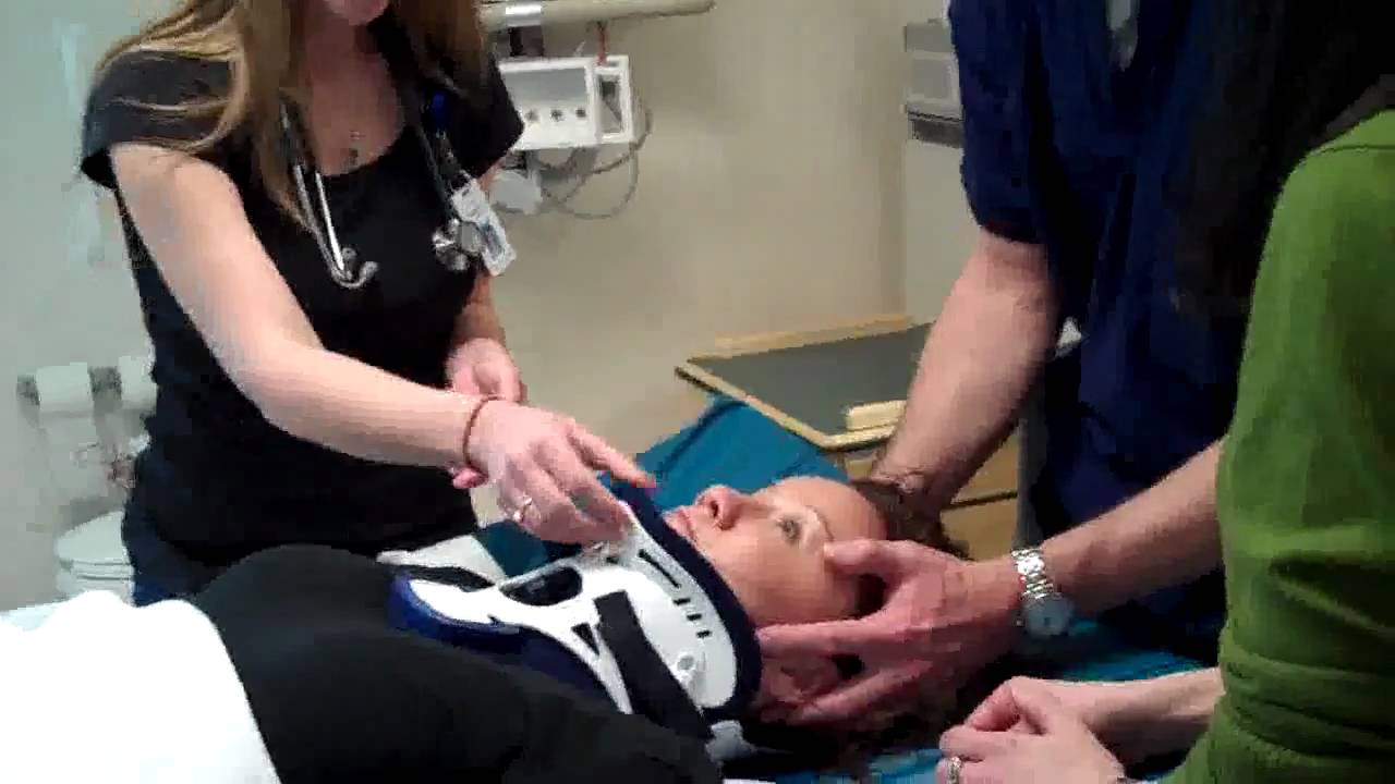

This video is for nurses, phsyicians, medics, and anyone else who has to put one of these on. Looks simple, but there are some nuances you need to know about.
Urgent and emergent intubation is challenging enough, but what if your patient is sporting some type of tongue piercing? Does it make a difference? Do you need to do anything differently?
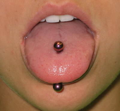
Obviously, the jewelry may physically impede the process of intubating the patient, impairing visualization of structures or getting in the way of inserting the tube. It can also cause complications later down the road, such as pressure necrosis from the tube coming into contact with it.
The anesthesia literature recommends removing all oral jewelry prior to elective intubation, or declining to do the case if the patient refuses. Unfortunately, trauma professionals do not have that option when the patient needs an emergency airway.
Here are some pointers for dealing with oral jewlry:
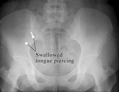
Note: both hands must always be in contact with the jewelry at all times! It is slippery, and if the pieces are not controlled, this can happen!
Chest tubes are needed occasionally to help manage chest injuries. How do you decide when they are ready for removal?
Unfortunately, the literature is not very helpful in answering this question. To come up with a uniform way of pulling them, our group looked at any existing literature and then filled in the blanks, negotiating criteria that we could all live with. We came up with the following.
Removal criteria:
Removal sequence:
Click here to download the full printed protocol.
Here’s a short video that shows you everything you need to know. Enjoy!
