Category Archives: Tips

How To MacGyver A Ventilator From Common Parts

A cardiac anesthesiology fellow, and several engineers from the University of Minnesota and a local device company got together over the past few weeks and cobbled together a ventilator from some spare parts. Here’s a picture to give you an idea of what it looks like:

It uses a metal toolbox tray, an Ambu bag, and some other spare parts lying around one of the medical device labs at the university. Essentially, a servo motor intermittently squeezes the Ambu bag, and there are adjustments for how often (rate) and how deeply (volume) the bag is compressed. There is a pressure limiting device included in the system as well.

This project illustrates how we will need to think outside the (tool)box in the coming weeks, especially as the number of severe Coronavirus cases begins to tax our supply of ventilators. And obviously, this thing will not get FDA approval in your lifetime. But if a choice needs to be made between using something like this in a pinch vs letting someone asphyxiate, the answer is pretty clear.

The group has produced a short YouTube video as well (see below), but it is rather short on details. You get to see some partial views of it as it is being tested on pigs. But so far, the concept is promising.

If you are trying to view this on my Tumblr feed, the video will not show here. Please click here to visit my main blog site to view it.

YouTube player
Print Friendly, PDF & Email

Coronavirus: Using One Ventilator To Ventilate Up To Four Patients

It’s unusual for me to post on a Saturday, but we are currently living in unusual times. This tidbit caught my attention and I wanted to get this interesting idea out there for all to think about.

Hopefully, it will never come to this. But there are reports from both Italy and New York City that ventilators are in short supply. This video illustrates the technique used in some research that was actually published way back in 2006. It demonstrated the efficacy of using one ventilator on four patients at once.  It requires just a few connectors that are readily available.

But remember, this is off-label use and is not condoned by the FDA! And it probably violates every Department of Health and hospital policy written. But in a pinch, it is something to think about.

Obviously, there are lots of possible downsides. Four patients are essentially sharing one circuit. Even though the circuits are one-way, there is always an infectious cross-contamination risk. The paper shows that ventilation should be adequate on both pressure and volume control settings.

This type of thing is a very last resort, and hopefully no one will have to use it!

YouTube player

Reference: A single ventilator for multiple simulated patients to meet disaster surge. Acad Emerg Med 13(11):1246-9, 2006.

Print Friendly, PDF & Email

Thoughts On Traumatic Hematuria: Part 2

Yesterday, I discussed blood in the urine from a urethra. As I mentioned, there is typically not much from that particular injury. Today, I’ll dig into the three causes of real hematuria.

All of these tubes show gross hematuria except the one on the right.

  • Bladder injury. This can occur with either blunt or penetrating injury. The degree of hematuria is variable with stabs or gunshots, but tends to be much darker in blunt injury. This happens because the size of the bladder injury tends to be greater with blunt force. The bladder injury is not necessarily full-thickness with blunt trauma. It may just be some wall contusion and underlying mucosal injury. But frequently, with seat belt injury and/or A-P compression injuries to the pelvis (“open book”), the injury is full thickness.
    • Tip: If less than 50cc of very dark urine flow from the catheter upon insertion, it is likely that your patient has an intraperitoneal bladder rupture!
  • Ureteral injury. This injury is very rare. The most common mechanism is penetrating, but this structure is so small and deep that it seldom gets hit by naything. Patients with multiple lumbar transverse process fractures will occasionally have a small amount of hematuria, probably from a minor contusion. More often than not, the hematuria is microscopic, so we should never know about it.
  • Kidney injury. The most important fact regarding renal injury is that the degree of injury has no correlation with the amount of hematuria. The most devastating injury, a devascularized kidney, frequently has little if any gross hematuria. And conversely, a very minor contusion can produce very red urine.

So what about diagnosis? It’s easy! If you see gross hematuria, insert a foley catheter (if not already done) and order a CT of the abdomen/pelvis with contrast, as well as a CT cystogram. The latter must not be done using passive filling of the bladder with a clamped catheter. Contrast must be infused into the bladder under pressure to ensure a bladder injury can be identified.

CT scan is an excellent tool for defining injuries to kidney, ureter, and bladder, and will identify extravasation into specific places and allow grading. Specific management will be the topic of future posts.

Print Friendly, PDF & Email

Thoughts On Traumatic Hematuria: Part 1

I’ve seen a number of patients recently with bloody urine, and that is prompting me to provide some (written) clarity to others who need to manage this clinical problem. I’ll try to keep it organized!

There are two kinds of hematuria in trauma: blood that you can see with the naked eye, and…

Okay, so there’s only one. Trauma professionals do not care about microscopic hematuria. It does not change clinical management. Sure, your patient might have a renal contusion, but you won’t do anything about that. Or, he/she might have an infarcting kidney. And you can’t do anything about that. If you order a urinalysis, you might see a few RBCs. Don’t let this lead you down the path of looking for a source. You’ll end up ordering lots of tests and additional imaging, and generally will have nothing to show for it at the end. It’s not your job to spend good money on the very rare chance of finding something clinically significant.

Both of these specimens have blood in them. You can’t see it on the left, so don’t go looking for it with a microscope.

There are four sources of blood in the urine.

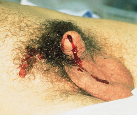
1. The first source does not generally cause hematuria, but can occasionally cause a few visible wisps of blood. That source is a urethral injury. The textbook teaching, and it’s good advice, is to look at the urethral meatus in your trauma patient, especially if you are contemplating insertion of a urinary catheter. If you see a few drops of blood, pause to consider. Sometimes, the blood is no longer visible, but might be present as a few well-placed drops on the patient’s underwear. So have a look at that, too, especially in patients with high risk injuries such as A-P compression pelvic fractures (think, lots of ramus fractures or pubic diastasis).

If you didn’t notice it and inserted the catheter anyway, you might see a few wisps of blood in the tubing as you place it. More often than not, this is just run of the mill irritation of the mucosa by the catheter, but always keep the possibility of an injury in mind.

Tomorrow, I’ll discuss the remaining three sources, and what to do about them.

Related posts:

Print Friendly, PDF & Email

Management Of Penetrating Neck Trauma: The Way We Were/Are

The management of penetrating injuries to the neck has changed very little over the years. Could it be time? Today, I’ll review some of the basics of classic diagnosis and treatment. In my next post, I’ll discuss an alternative way to approach it.

First, lets look at the time-honored zones of the neck. Here’s a nice diagram from EMDocs.net:

The zones are numbered in reverse, from bottom to top, and in Roman numerals.

The area below the cricoid cartilage is considered Zone I and contains many large vascular and aerodigestive structures that are relatively difficult to approach surgically. For this reason, diagnostic testing is recommended to assist in determining if an operation is actually needed and what the best surgical exposure would be. Obviously, this can only be considered in the stable patient. Unstable patients must go straight to the OR and the trauma surgeon will determine the surgical approach on the fly.

Similarly, the area above the angle of the mandible is Zone III, and is also difficult to expose. Injuries to this area may involve the distal carotid and vertebral arteries near the base of the skull, as well as the distal jugular vein. Surgical approach may require dislocation of or fracturing the mandible to get at this area. This is  challenging and not that desirable, and few surgeons are familiar with the technique. For this reason, imaging is very desirable and often demonstrates that no significant injury is present. And endovascular / angiographic techniques are now available that may obviate the need for surgery.

Zone II is everything in-between the mandibular angle and cricoid cartilage. This is the surgical Easy Button. Exposure is simple and the operation is fun. In the old days, an injury to this area went straight to the OR regardless of whether there were signs or symptoms of injury. Yes, there were quite a few negative explorations. But we’ve become more selective now with the advent of improved resolution of our CT scans.

Currently, we usually follow a two-step approach to penetrating neck trauma:

  1. Are there hard signs of injury present? These tell us that a structure that absolutely needs to be fixed has been injured. The patient should be taken directly to OR after control of the airway, if appropriate. Typical hard signs are:
    1. Airway compromise
    2. Active air bubbling from wound
    3. Expanding or pulsatile hematoma
    4. Active bleeding
    5. Hematemesis
  2. What zone is the injury in? And don’t just look at the obvious entry point. Gunshots (and long knives) may enter multiple zones. The zone then determines what happens next:
    1. Zone I – CT angio of neck and chest. If positive, proceed to OR for repairs, and perform EGD and/or bronchoscopy as needed
    2. Zone II – Old days: proceed to operating room for exploration, or angiogram, EGD, direct laryngoscopy, and bronchoscopy. Most chief residents chose the former. Current day: CTA of neck, followed by OR, EGD, bronchoscopy only if indicated.
    3. Zone III – CT angio of the neck. If positive, consider angiography/endovascular consultation vs operation.

Changes from old days to more current thinking have been made possible by improvements in speed and resolution of our CT scanners. But why can’t we take this another step forward and streamline this process even more? I’ll propose some changes in my next post!

Reference: Western Trauma Association Critical Decisions in Trauma:
Penetrating neck trauma. J Trauma 75(6):936-940, 2013.

Print Friendly, PDF & Email