Category Archives: Tips

Management Of Penetrating Neck Trauma: The Way We Were/Are

The management of penetrating injuries to the neck has changed very little over the years. Could it be time? Today, I’ll review some of the basics of classic diagnosis and treatment. In my next post, I’ll discuss an alternative way to approach it.

First, lets look at the time-honored zones of the neck. Here’s a nice diagram from EMDocs.net:

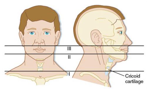
The zones are numbered in reverse, from bottom to top, and in Roman numerals.

The area below the cricoid cartilage is considered Zone I and contains many large vascular and aerodigestive structures that are relatively difficult to approach surgically. For this reason, diagnostic testing is recommended to assist in determining if an operation is actually needed and what the best surgical exposure would be. Obviously, this can only be considered in the stable patient. Unstable patients must go straight to the OR and the trauma surgeon will determine the surgical approach on the fly.

Similarly, the area above the angle of the mandible is Zone III, and is also difficult to expose. Injuries to this area may involve the distal carotid and vertebral arteries near the base of the skull, as well as the distal jugular vein. Surgical approach may require dislocation of or fracturing the mandible to get at this area. This is  challenging and not that desirable, and few surgeons are familiar with the technique. For this reason, imaging is very desirable and often demonstrates that no significant injury is present. And endovascular / angiographic techniques are now available that may obviate the need for surgery.

Zone II is everything in-between the mandibular angle and cricoid cartilage. This is the surgical Easy Button. Exposure is simple and the operation is fun. In the old days, an injury to this area went straight to the OR regardless of whether there were signs or symptoms of injury. Yes, there were quite a few negative explorations. But we’ve become more selective now with the advent of improved resolution of our CT scans.

Currently, we usually follow a two-step approach to penetrating neck trauma:

  1. Are there hard signs of injury present? These tell us that a structure that absolutely needs to be fixed has been injured. The patient should be taken directly to OR after control of the airway, if appropriate. Typical hard signs are:
    1. Airway compromise
    2. Active air bubbling from wound
    3. Expanding or pulsatile hematoma
    4. Active bleeding
    5. Hematemesis
  2. What zone is the injury in? And don’t just look at the obvious entry point. Gunshots (and long knives) may enter multiple zones. The zone then determines what happens next:
    1. Zone I – CT angio of neck and chest. If positive, proceed to OR for repairs, and perform EGD and/or bronchoscopy as needed
    2. Zone II – Old days: proceed to operating room for exploration, or angiogram, EGD, direct laryngoscopy, and bronchoscopy. Most chief residents chose the former. Current day: CTA of neck, followed by OR, EGD, bronchoscopy only if indicated.
    3. Zone III – CT angio of the neck. If positive, consider angiography/endovascular consultation vs operation.

Changes from old days to more current thinking have been made possible by improvements in speed and resolution of our CT scanners. But why can’t we take this another step forward and streamline this process even more? I’ll propose some changes in my next post!

Reference: Western Trauma Association Critical Decisions in Trauma:
Penetrating neck trauma. J Trauma 75(6):936-940, 2013.

Print Friendly, PDF & Email

Trauma Tip: The “Dang!” Factor

This issue continues to rear its ugly head, so I continue to repost from time to time.

This tip is for all trauma professionals: prehospital, doctors, nurses, etc. Anyone who touches a trauma patient. You’ve probably seen this phenomenon in action. A patient sustains a very disfiguring injury. It could be a mangled extremity, a shotgun blast to the torso, or some really severe facial trauma. People cluster around the injured part and say “Dang! That looks really bad!”

It’s just human nature. We are drawn to extremes, and that goes for trauma care as well. And it doesn’t matter what your level of training or expertise, we are all susceptible to it. The problem is that we get so engrossed (!) in the disfiguring injury that we ignore the fact that the patient is turning blue. Or bleeding to death from a small puncture wound somewhere else. We forget to focus on the other life threatening things that may be going on.

How do we avoid this common pitfall? It takes a little forethought and mental preparation. Here’s what to do:

  • If you know in advance that one of these injuries is present, prepare your crew or team. Tell them what to expect so they can guard against this phenomenon.
  • Quickly assess to see if it is life threatening. If it bleeds or sucks, it needs immediate attention. Take care of it immediately.
  • If it’s not life threatening, cover it up and focus on the usual priorities (a la ATLS, for example).
  • When it’s time to address the injury in the usual order of things, uncover, assess and treat.

Don’t get caught off guard! Just being aware of this common pitfall can save you and your patient!

Print Friendly, PDF & Email

Mistaken Identity In Trauma Care

There was a well-publicized and tragic case of mistaken identity after a motor vehicle crash in Indiana a number of years ago. A van carrying several college students and staff crashed, resulting in multiple fatalities at the scene. Survivors were transported to a Michigan trauma center, and it wasn’t until five weeks later that the identity mixup was discovered.

One of the fatally injured students and one of the survivors were both female, blonde, and about the same height and size. Their identities were not confirmed because the next of kin of the deceased was advised not to look at the body. And the face of the surviving woman was significantly contused and she had sustained multiple facial fractures. She remained comatose and intubated for over month after the other was buried (by the wrong family, it turns out). After extubation, she began correcting people who called her by the deceased woman’s name, and the correct identification was finally made.

How can this happen?! It’s not as difficult as it might seem, for a number of reasons:

  • Faces and identifying marks may be mutilated
  • Position in the vehicle may be mistaken
  • Bystander descriptions are notoriously inaccurate in these situations

It is neither practical nor safe to delay transport from the scene in the interest of obtaining positive identification. And hospitals have even less information than prehospital providers, whom they rely on almost exclusively for accurate data.

What can be done to avoid a case of mistaken identity? EMS and hospitals must develop protocols to follow in any case where multiple patients are treated at once. The baseline assumption must be that the identities are unclear or unknown until definitively made, and preferably from multiple sources. What are these definitive items?

  • An official ID that is still on the victim’s person (not cut off in the clothes)
  • Self identification
  • Visual identification from someone who personally knows the victim and views or talks to them
  • Written description, where the patients have very different identifying characteristics

However, remember that every one of these can be made in error. This is why multiple sources are so important. If in doubt, the patients should remain a “Doe” and not be given a real name.

If you have specific protocols or policies, please share them with me by email so I can post them!

Print Friendly, PDF & Email

Best Practice: Laundry Basket In The Resus Room?

How do you get patients out of their clothes during a trauma resuscitation? Most of the time, I bet your answer is “with a pair of scissors.” And once they are off, what do you do with them? Admit it. You just throw them on the floor. And sometime later, someone’s job is to find it all, put it in a bag, and store it or hand it over to the police.

There are more problems than you might think with this approach. First, and most importantly to the patient, their stuff can get lost. Swept up with all the other detritus from a trauma activation. And second, their belongings may become evidence and it’s just been contaminated.

So here’s an easy solution. Create a specific place to put the clothes. Make it small, with a tiny footprint in your trauma room. Make it movable so it can be kept out of the way. And make sure it is shaped so it can contain a large paper bag to preserve evidence without contamination.

And here’s the answer:

Yes, it’s a plain old laundry basket. The perfect solution. And best of all, these are dirt cheap when you are used to seeing what hospitals charge for stuff. So your ED can buy several ($14.29 ea on Amazon.com) in case they can’t be cleaned anymore or just disappear.

Print Friendly, PDF & Email

Next Week: The Electronic Trauma Flow Sheet

It’s been a while since I visited the topic of the electronic trauma flow sheet (eTFS) for trauma activations. A few years have passed, the software engineers have gotten smarter (hopefully), so let’s look at it again.

Next week, I’ll be covering the eTFS in detail again. Here is what I will touch on:

  • eTFS: Why Hospitals Want To Switch
  • What Does(n’t) Work
  • Oops! Now What Are My Options?

I’d also like to spend a day on Q&A. To that end, please start emailing, commenting, or tweeting your questions so I can answer them in detail at the end of the week. Let the games begin!

Print Friendly, PDF & Email