Category Archives: Solid organ

Best of AAST #12: Embolization Of Splenic Pseudoaneurysm

The management of blunt spleen injury has evolved significant over the time I’ve been in practice. Initially, the usual formula was:

Spleen injury = splenectomy

This began to change in the late 1980’s, and beginning in the early 90’s nonoperative management became the rage. We spent the next 10-15 years tweaking the details, gradually reducing bed rest and NPO times, and increasing the success rate through smart patient selection and discovering new adjuncts.

One of these adjuncts was angiography with embolization. The ShockTrauma Center in Maryland was an early adopter and protocolized its use in patients with high-grade injuries.

But now, they are questioning the utility of this tool in certain patients: those with splenic pseudoaneurysms (PSA). They theorized that modern, high resolution CT identifies relatively unimportant pseudoaneurysms. They conducted a 5-year retrospective review of their experience.

Here are the factoids:

  • They identified 717 splenic injuries, of whom 155 were embolized but only 140 patients had adequate records and imaging for review
  • The majority of patients had high grade injury: 31% Grade 3, 61% Grade 4, 1% Grade 5
  • Extravasation was seen in 17% and PSA in 52%
  • About 44% of patients went to angiography within 6 hours, but the mean was 17 hours indicating quite a few outliers
  • Among the 73 patients with an initial PSA , a third of them did not have a detectable lesion during angiography
  • Patients who underwent embolization for PSA had a followup CT 48-72 hours afterwards, persistently perfused PSA were seen in 40% (!)
  • No patients with PSA who were only observed required delayed splenectomy

The authors conclude that a third of pseudoaneurysms may be clinically insignificant, and that 40% of them persist after embolization. They do not, however, offer any recommendations based on their data.

Here are my comments: This is an interesting study. My read of the abstract and slides would indicate that this group routinely sends all Grade 3 and 4 injuries to angio, and Grade 5 could go to either angio or OR. They take their good time going to interventional radiology (mean 17 hours from arrival), and get a routine followup CT 48-72 hours from hitting the door if they didn’t go to the OR.

If I were to play the devil’s advocate, I might think that interventional radiology was being de-emphasized for some reason. Was there some reluctance to send patients there, or limited availability? This might explain the long access times. And how are the radiologists not shutting down 40% of PSA that are seen?

I am intrigued by the study, but there are a lot more details needed to get some good takeaways from it.

Here are my questions for the presenter and authors:

  • Please explain why it takes so long to send patients to angiography. Less than half got there in less than 6 hours, and the mean of 17 hours means that many didn’t get there until the next day.
  • Does this small study have the statistical power to say that some PSA are benign? The groups are very small, and I would speculate that the group size needed to show significance is in the high hundreds.
  • What was the reason for splenectomy in the 2 patients who underwent embolization? Was it related to the pseudoaneurysm or something else?
  • How can you be sure that these PSA are insignificant? Frequently, pseudoaneurysms don’t explode for 7-10 days. Do you have any data on patients who returned to a hospital with delayed bleeding?
  • If you believe that many pseudoaneurysms are benign, how do you propose to manage the patients? Observe until they explode? Repeat a contrast CT scan, with the associated contrast and radiation re-dose? And how long would you wait to do this? What would your new protocol be?

I’ll be all ears on Friday when this abstract is presented live.

Serial Hemoglobin / Hematocrit – Huh? Part 2

In my last post, I waxed theoretical. I discussed the potential reasons for measuring serial hemoglobin or hematocrit levels, the limitations due to the rate of change of the values, and conjectured about how often they really should be drawn.

And now, how about something more practical? How about an some actual research? One of the more common situations for ordering serial hemoglobin draws occurs in managing solid organ injury. The vast majority of the practice guidelines I’ve seen call for repeating blood draws about every six hours. The trauma group at the University of Florida in Jacksonville decided to review their experience in patients with liver and spleen injuries. Their hypothesis was that hemodynamic changes would more likely change management than would lab value changes.

They performed a retrospective review of their experience with these patients over a one year period. Patients with higher grade solid organ injury (Grades III, IV, V), either isolated or in combination with other trauma, were included. Patients on anticoagulants or anti-platelet agents, as well as those who were hemodynamically unstable and were immediately operated on, were excluded.

Here are the factoids:

  • A total of 138 patients were included, and were separated into a group who required an urgent or unplanned intervention (35), and a group who did not (103)
  • The intervention group had a higher ISS (27 vs 22), and their solid organ injury was about 1.5 grades higher
  • Initial Hgb levels were the same for the two groups (13 for intervention group vs 12)
  • The number of blood draws was the same for the two groups (10 vs 9), as was the mean decrease in Hgb (3.7 vs 3.5 gm/dl)
  • Only the grade of spleen laceration predicted the need for an urgent procedure, not the decrease in Hgb

Bottom line: This is an elegant little study that examined the utility of serial hemoglobin draws on determining more aggressive interventions in solid organ injury patients. First, recognize that this is a single-institution, retrospective study. This just makes it a bit harder to get good results. But the authors took the time to do a power analysis, to ensure enough patients were enrolled so they could detect a 20% difference in their outcomes (intervention vs no intervention). 

Basically, they found that everyone’s Hgb started out about the same and drifted downwards to the same degree. But the group that required intervention was defined by the severity of the solid organ injury, not by any change in Hgb.

I’ve been preaching this concept for more than 20 years. I remember hovering over a patient with a high-grade spleen injury in whom I had just sent off the requisite q6 hour Hgb as he became hemodynamically unstable. Once I finished the laparotomy, I had a chance to pull up that result: 11gm/dl! 

Humans bleed whole blood. It takes a finite amount of time to pull fluid out of the interstitium to “refill the tank” and dilute out the Hgb value. For this reason, hemodynamics will always trump hemoglobin levels for making decisions regarding further intervention. So why get them?

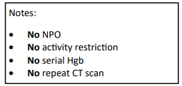
Have a look at the Regions Hospital solid organ injury protocol using the link below. It has not included serial hemoglobin levels for 18 years, which was when it was written. Take care to look at the little NO box on the left side of the page.

I’d love to hear from any of you who have also abandoned this little remnant of the past. Unfortunately, I think you are in the minority!

Click here for the Regions Hospital Solid Organ Injury Protocol

Reference: Serial hemoglobin monitoring in adult patients with blunt solid organ injury: less is more. J Trauma Acute Care Open 5:3000446, 2020.

Update: Kidney Injury Scaling

Over the past two days, I’ve reviewed the new AAST organ injury scaling updates for spleen and liver injuries. Today, I’ll cover the new kidney grading scale.

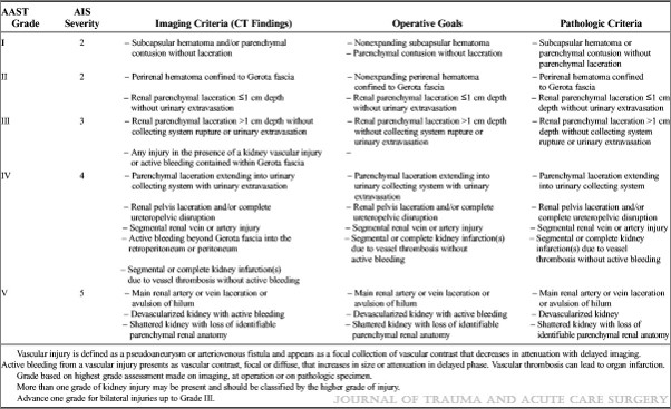
Liver and spleen grading is generally simple, focusing on laceration depth and subcapsular hematoma coverage to determine the exact value. However, the kidney is totally different. Although technically a solid organ, it’s got a bunch of hollow, urine-containing stuff inside. This is the main determinant of the original scaling system: collection system involvement.

Like liver and spleen, the kidney scale was updated to take advantage of CT information. But once again, bleeding identified via the CT angiogram is incorporated into the higher grades. Active bleeding contained within Gerota’s fascia is assigned a grade of III. Extravasation escaping this fascia is assigned a IV.  The other grades remain unchanged.

Here are the updated guidelines. Click the image or link below it to open a bigger image in a new window.

Click to download larger image

Links:

Update: Liver Injury Scaling

In my last post, I reviewed the updated AAST organ injury scaling (OIS) for the spleen. Today, I’ll share details of the new version of liver grading.

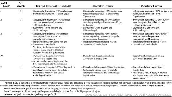
First, the overall focus of the updated liver scale is similar to the spleen one: it incorporates a listing of criteria identified by CT scan that parallels the old anatomic criteria. The CT column contains all the old anatomic stuff, but now includes scaling for active bleeding.

The confusing part? Whereas contained active bleeding within the spleen was Grade IV and active bleeding escaping the spleen was Grade V in the updated scale, these drop down a grade in the liver. So bleeding contained with the liver parenchyma is Grade III and active extravasation escaping into the peritoneal cavity is only Grade IV. I presume this has to do with the abbreviated injury score (AIS) used to calculate ISS, and that the mortality hit from this degree of bleeding is less than that of the spleen.

The final difference between the updated scale and the original is the removal of Grade VI. This was previously described as hepatic avulsion, which is a nonsurvivable injury. The AIS for Grade VI liver used to be 6, which causes an immediate ISS calculation short circuit to 75. Which also means that survival is approximately 0%. This is not part of the OIS update, which may be due to the fact that it never occurs in anyone who makes it to a trauma center alive.

Here are the updated guidelines. Click the image or link below it to open a bigger image in a new window.

Click to download larger image

In the next post, I’ll review the new features of the kidney injury scale.

Update: Spleen Injury Scaling

Over the years, the American Association for the Surgery of Trauma (AAST) has developed and maintained a library of organ injury scales. Organ injury scaling allows us to compare apples to apples in research studies, and in many cases enables us to tailor interventions and predict outcomes. Many of the scales have been in place for decades and have not been updated. The spleen, liver, and kidney scales were introduced 25 year ago, and received their first update last December. During the next three posts, I’ll review what’s new and different with them.

The biggest change to all three scales has been the incorporation of specific vascular injuries seen on modern-day CT scans. It is recommended that scanning for solid organ injury be conducted using dual phase (arterial and portal venous) scanning techniques. This increases study sensitivity and provides the best images for accurate diagnosis and scaling. Also note that specific criteria are now provided for CT, intraoperative, and pathologic diagnosis.

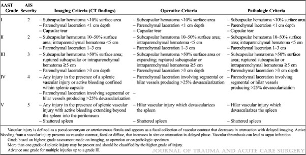
Let’s start with the spleen today. Here are the updated guidelines. Click the image or link to get a bigger image in a new window.

Click to download larger image

The main change to this scale is the addition of active bleeding contained within the spleen (pseudo-aneurysm or contained extravasation) to Grade IV, and uncontained extravasation to Grade V.

In my next post, I’ll discuss the new features of the liver injury scale.