Category Archives: Performance Improvement

Best Of AAST 2021: Reducing Errors In Trauma Care

Finally, a performance improvement (PI) abstract at AAST!

As many of you know, there are two general types of issues that are encountered in the usual PI processes: provider (peer) vs system. Provider issues are errors of omission or commission by an individual clinician. Examples include a surgeon making a technical error during a procedure, or prescribing the wrong drug or dose for some condition.

One might think that provider issues are the most common type of problem encountered. But they would be wrong. The vast majority of clinicians go to work each day with the idea that they will do their job to the best of their abilities. So how could things go awry?

Because the majority of errors have some degree of system component! They are set up to fail by factors outside their perception and/or control. Let’s look at a surgeon who has several small bowel anastomoses fall apart. His surgery department head chides/educates him, reports him to hospital quality, and proctors his next ten bowel cases. Everything is good, right?

But then, two months later, the stapler company issues a recall because they found a higher than usual number of anastomotic failures with one of their products. So it wasn’t the surgeon after all, like everyone assumed. This is an extreme example, but you get the idea. System issues often look like peer issues, but it’s frequently difficult for many PI programs to recognize or accept this.

A multi-institutional group reviewed the results of a newly implemented Mortality Reporting System (MRS) to analyze a large number of PI opportunities for improvement (OFI). More than 300 trauma centers submitted data to the MRS when a death occurred where an OFI was identified. The reports included details of the incident and mitigation strategies that were applied.


Here are the factoids:

  • A total of 395 deaths were reviewed over a two year period
  • One third of deaths were unanticipated (!!), and a third of those were failure to rescue
  • Half of errors pertained to clinical management, clinical performance, and communication
  • Human failures occurred in about two thirds of cases
  • The most common remedy applied was education, which presumes a “provider issue”
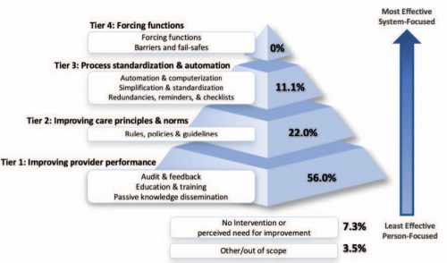
  • System strategies like automation, standardization, and fail-safe approaches were seldom used, implying that system issues were seldom recognized
  • in 7%, the trauma centers could not identify a specific strategy to prevent future harm (!!!)

The authors concluded that most strategies to reduce errors focus on individual performance and do not recognize the value of system-level intervention.

Bottom line: Look at the pyramid chart above (interesting choice for a chart, but very effective). The arrow shows progression from provider focus to systems focus. The pyramid shows how the recognition of and intervention for system issues drops off very rapidly.

I am both shocked and fascinated by the last bullet point. A strategy couldn’t be developed to prevent the same thing from happening again. Now, there are a few rare instances where this could be correct. Your patient could have been struck by a bolt of lightning in her room, or a meteorite could have crashed through the wall. But I doubt it. This 7% illustrates the importance of investigating all the angles to try to determine how the system failed!

For once, I have no critique for an abstract. It is a straightforward descriptive study that reveals an issue that many in PI are not fully aware of. I’ll definitely be listening to this one, and I really look forward to the published paper!

Reference: ERROR REDUCTION IN TRAUMA CARE: LESSONS FROM AN ANONYMIZED, NATIONAL, MULTI-CENTER MORTALITY REPORTING SYSTEM. AAST 2021, Oral abstract #17.

Print Friendly, PDF & Email

Trauma Performance Improvement: How Often Should Your Committees Meet?

For those of you who are trauma performance aficionados, check out the newest posts on my PI site TraumaMedEd.com. There is a post for each of your required trauma program committees:

How Often Should My Multidisciplinary Trauma PI Committee Meet?

and

How Often Should My Trauma Operations Committee Meet?

And if you are interested in receiving email notices when new PI-related posts are available, please subscribe by clicking here!

Print Friendly, PDF & Email

Mainstem Intubation In Pediatric Patients: How To Avoid It

In my last post, I reviewed a simple technique utilizing a reminder card and provider feedback loops to reduce deep intubations in pediatric patients. Today, I’ll review three other techniques and discuss a paper that compared their efficacy.

A variety of techniques for determining and/or confirming endotracheal tube position exist. Use of one or more of them is important in children due to their short trachea and increased likelihood of deep intubation. Some, like the confirmatory chest x-ray, are obvious. However, it’s more desirable to apply techniques during the intubation in order to avoid deep intubation in the first place. Hyperinflation of one lung, especially in very small children, can cause a host of impairments and complications that may compound their other injuries.

A paper from the University Hospital Basel in Switzerland evaluated three techniques: bronchoscopic insertion to a specific depth, cuff palpation in the sternal notch, and intentional right mainstem intubation followed by slow withdrawal during auscultation.  Each of 68 children ranging in age from 0 to 4 years were studied using all three techniques.

Each endotracheal tube was marked at the ideal insertion point that would ideally be placed just beyond the vocal cords. The distance from this mark to the mouth end of the tube was measured so actual intubation depths could be compared.

Bronchoscopic insertion was always performed first to obtain a baseline depth measurement, essentially the gold standard. The other two techniques were performed in random order. For the cuff palpation technique, the trachea was palpated while the balloon was intermittently partially inflated until it could be felt at the suprasternal notch. For the mainstem intubation technique, the uninflated tube was advanced until breath sounds in the left axilla disappeared. It was then slowly withdrawn until sounds reappeared.

Distances from the tip of the tube to the carina was calculated using the insertion depth at the incisors and the initial ideal intubation depth mark. Here are the factoids:

  • Insertion to a depth mark on the tube via bronchoscope technique resulted in the highest tube tip with respect to the carina, and also with the greatest depth variability
  • The cuff palpation technique resulted in less distance to the carina (about 19mm vs 36 for the mark technique) and less variability
  • Use of the mainstem intubation with pullback technique resulted in the tube tip resting within just a few mm of the carina, but tube depth was very consistent

Bottom line: What to make of all this? Which technique is “best?” First, it’s not practical or advisable to use a bronchoscope for every pediatric intubation. It’s invasive and adds complexity and time to a critical procedure. The cuff palpation technique also takes additional time due to the repeated cuff inflation/deflation that is required. However, the tube position is fairly accurate and safe.

The intentional right mainstem intubation with pull-back seems a bit sketchy. It requires some type of ongoing ventilation while the tube is being inserted, as well as someone who can listen to the left chest. Additionally, it results in a tube position that is so low that neck positioning may move it into the mainstem bronchus again.

In my mind, estimation of the proper depth pre-intubation is probably the best. Strict attention must be paid to the final depth of the tube once it is inserted, as measured by the distance marker at the incisors. This number must match the one decided upon at the start of the procedure. A good exam of the chest should be carried out to quickly identify an inadvertent mainstem intubation. And finally, a quick confirmatory chest x-ray should always be obtained for objective information on tube position. 

The fancy techniques described in this paper add too much time and complexity for intubation in a trauma situation. They may very well have a place in the OR where the situation is more controlled and there is more advanced equipment and support. But stick to the basics when intubating children in your trauma bay!

Reference: Assessment of three placement techniques for individualized positioning of the tip of the tracheal tube in children under the age of 4 years. Ped Anesthesia 25:379-285, 2014.

Print Friendly, PDF & Email

Mainstem Intubation In Pediatric Patients: How Common?

Mainstem intubation in the pediatric patient is a common problem. There are two major issues: the trachea is shorter than in an adult, and the angles are different making intubation of the right mainstem bronchus much easier. Frequently, the intubator watches the balloon slide between the cords, then pushes the tube in “just a little further.”

Unfortunately, that “little bit” can vary significantly. An abstract from my hospital was presented at the Pediatric Trauma Society in 2016. Subjectively, we noticed that mainstem intubation was occurring with some regularity in our pediatric trauma patients.  It seemed as though insufficient attention was being paid to the depth of the tube.

A major difference between adult and pediatric intubations is that in adults, optimal tube depth is locked into a relatively narrow range. In children, the depth varies considerably based upon child age and size. And small variances in depth can have major implications for tube position.

We decided to implement a PI project to change our intubation policy. In order to focus the entire team on tube depth, a color coded card was attached to each size of endotracheal tube. This card listed the optimal depth for insertion. Once the provider inserted the tube, the final depth was called out for the team and documentation scribe to hear. This had the added advantage of allowing multiple team members confirm the appropriateness of tube depth. A chest x-ray was immediately obtained to confirm position.

We retrospectively reviewed our seven year experience with pediatric intubations, from 2009-2015. Here are the factoids:

  • Nearly 2,000 pediatric trauma patients were admitted during the study period
  • 94 patients (5%) required intubation in the ED
  • Prior to implementation of the new protocol, 6 of 68 patients (8.8%) had confirmed right mainstem intubation 
  • After the change, only one further mainstem intubation occurred in 26 procedures (3.8%)

Bottom line: Unfortunately, this series is too small to determine statistical significance. There is a definite trend toward fewer mainstem intubations. It appears that by calling more attention to the proper tube depth, fewer deep placements occur. Our numbers  have remained low since this change.

Are there other methods to ensure proper ET tube placement in small patients? In my next post, I’ll review a paper that compares three additional different techniques that can be used.

Reference: Eliminating the Preventable Occurrence of Right Mainstem Intubation in the Pediatric Trauma Patient: A Quality Performance Improvement (PI) Initiative. Pediatric Trauma Society Poster Abstract #1, 2016.

Print Friendly, PDF & Email

What’s The Best Trigger For Your Massive Transfusion Protocol?

Every trauma center verified by the American College of Surgeons Committee on Trauma (ACS-COT) must have a massive transfusion protocol (MTP). The details and logistics of the protocol are up to the individual center. The difficult question is: how is a trauma professional to know that the MTP should be activated?

Sometimes it’s extremely obvious. The patient is very hypotensive. Blood is spurting all over the room. But sometimes it’s more subtle and the need just seems to creep up on you. And frequently, this delays activation and the actual arrival of the blood that is so desperately needed.

I’ve previously written about common triggers for the MTP, including psychic powers, shock index, and ABC index. See the links below to read my MTP week posts. But is one better than the other? The group at Vancouver General Hospital in British Columbia, Canada performed a systematic review of the literature to try to answer this question.

A total of 45 pertinent articles were identified in the literature up to 2017. Fifteen different scoring systems were evaluated involving combinations of clinical assessment, laboratory tests, and ultrasound evaluation.

Here are the factoids:

  • The best validated score using clinical assessment plus ultrasound was the Assessment of Blood Consumption score (click here for my post). This was the easiest to score compared to other systems using ultrasound.
  • Shock index (SI) was the only validated system using just the clinical exam
  • Some other studies were promising, with excellent areas under the receiver operating characteristic curve (AUROC), but had not been validated. The best of the bunch was one from Mina et al, but it is complicated enough to require a smartphone tool for calculation.
  • Other promising studies required laboratory evaluations which preclude their use at the time of patient arrival
  • Scoring systems that used more variables generally showed better correlation with actual need for MTP, but were more less likely to provide suficiently early predictions
  • Most validation studies involved single centers
  • No studies were designed to or able to show improved outcomes

Bottom line: There are many, many systems out there for predicting need for activation of the MTP (at least 15 to date)! This review concludes that the system used should be tailored to the center implementing it.

Simpler is better. I still recommend either Shock Index (SI) or ABC. Shock index is quickly calculated based on physical exam as heart rate divided by systolic blood pressure. The normal range is 0.5 to 0.7. The likelihood of MTP escalates 2x with SI > 0.9, 4x if SI > 1.1, and 7x with SI > 1.3. The ratio can easily be calculated based on numbers available from EMS providers prior to arrival. Basically, pick your threshold.

The Assessment of Blood Consumption (ABC) uses four parameters, three of which could be reported prior to patient arrival:

  • Heart rate > 120
  • Systolic blood pressure < 90
  • FAST positive
  • Penetrating mechanism

If two or more criteria are met, the patient has a 41% likelihood of needing MTP.

So basically, use a system that works for you. From my experience, centers that use a system tend to use ABC. But definitely pick a system, don’t leave it up to chance with the trauma surgeon. And use your trauma PI program to assess utilization to see if it’s the best tool for your center.

Related posts:

Reference: Systematic Reviews of Scores and Predictors to Trigger Activation of Massive Transfusion Protocols. Accepted ahead of print, J Trauma, 2019.

Print Friendly, PDF & Email