Category Archives: New technology

New Technology: Using AI To Interpret Pelvic X-rays

Look out, radiologists! The computers are coming for you!

Radiologists use their extensive understanding of human anatomy and combine it with subtle findings they see on x-ray shadow pictures. In doing this, they can identify a wide variety of diseases, anomalies, and injuries. But as we have seen with vision systems and game playing (think chess), computers are getting pretty good at doing this as well.

Is it only a matter of time until computer artificial intelligence (AI) starts reading x-rays?  Look at how good they already are at interpreting EKGs. The trauma group at Stanford paired up with the Chang Gung Memorial Hospital in Taiwan to test the use of AI for interpreting images to identify a specific set of common pelvic fractures.

The Stanford group used a deep learning neural network (XCeption) to analyze source x-rays (standard A-P pelvis images) from Chang Gung. These x-rays were divided into training and testing cohorts. The authors also applied different degrees of blurring, brightness, rotation, and contrast adjustment to the training set in order to help the AI overcome these issues when interpreting novel images.

The AI interpreted the test images with a very high degree of sensitivity, specificity, accuracy, and predictive values, with all of them over 0.90. The algorithms generated a “heat map” that showed the areas that were suspicious for fracture. Here are some examples with the original x-ray on the left and the heat map on the right:
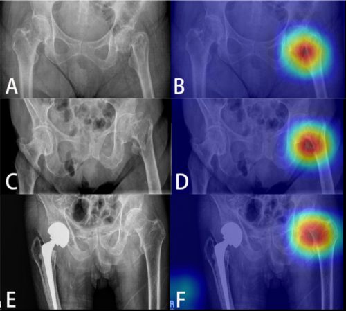
The top row shows a femoral neck fracture, the middle row an intertrochanteric fracture, and the bottom row another femoral neck fracture with a contralateral implant. All were handily identified by the AI.

AI applications are usually only as good as their training sets. In general, the bigger the better so they can gain a broader experience for more accurate interpretation. So it is possible that uncommon, subtle fractures could be missed. But remember, artificial intelligence is meant to supplement the radiologist, not replace him or her. You can all breathe more easily now.

This technology has the potential for broader use in radiographic interpretation. In my mind, the best way to use it is to first let the radiologist read the images as they usually do. Once they have done this, then turn on the heat map so they can see any additional anomalies the AI has found. They can then use this information to supplement the initial interpretation.

Expect to see more work like this in the future. I predict that, ultimately, the picture archiving and communications systems (PACS) software providers will build this into their product. As the digital images are moving from the imaging hardware to the digital storage media, the AI can intercept it and begin the augmented interpretation process. The radiologist will then be able to turn on the heat map as soon as the images arrive on their workstation.

Stay tuned! I’m sure there is more like this to come!

Reference: Practical computer vision application to detect hip fractures on pelvic X-rays: a bi-institutional study.  Trauma Surgery and Acute Care Open 6(1), http://dx.doi.org/10.1136/tsaco-2021-000705.

When Did The Surgeon Arrive At The Trauma Activation?

All trauma centers have mandatory arrival requirements for the surgeon at their highest-level trauma activations. Most Level I and II centers abide by the American College of Surgeons (ACS) requirement of 15 minutes after patient arrival. Level III centers typically mandate 30 minutes for their highest-level activation. And failure to meet these criteria can actually lead to loss of verification.

But what is the best way to record this critical piece of information? A number of methods have been used over the years. The earliest was simply recording the time of surgeon arrival on the paper trauma flow sheet. This has evolved over the years as technology has advanced. Most hospitals have installed badge swipe systems, since name badges have become nearly ubiquitous for gaining access to restricted areas within the hospital. A paper published last year details one hospital’s experience using a badge swipe system to do just this.

A NYC metro area Level I center started using a name badge swipe system to record the surgeon’s arrival in the ED for trauma activations several years ago. They examined their trauma activation data over a 7 month period at the end of 2016. Surgeon arrival times were recorded on the trauma flow sheet, and the electronic swipe information was included to supplement flow sheet results.

Here are the factoids:

  • There were 531 trauma activations during the study period, with 50 highest-level activations and 481 limited activations
  • The overall paper trauma flow sheet completion rate was 50% without card swipe data (!!)
  • For highest-level activations, surgeon presence was documented in 76%, but they arrived on time (< 15 minutes) only 70% of the time (!!!)
  • For intermediate-level activations, surgeon arrival was recorded 47% of the time and the surgeon was on time 45% of the time (I’m running out of exclamation points!!)
  • After including the badge swipe data, overall completion rate “improved” to 70%, which broke down to 90% in highest-level and 68% in the intermediate level activations
  • Surgeon compliance with arrival times improved to 84% and 63% for the two activation levels

The authors blamed the poor record keeping and compliance on “the fast pace of an ED.” They concluded that the badge swipe system was successful in increasing documentation and arrival compliance.

Bottom line: Oh, this is a fail on so many levels! First, surgeon arrival timeliness was appalling both with and without the badge swipe data. It started at 50% and increased to a barely passing score of 84%. And since this center only receives 100 highest-level activations per year, just a few more slip-ups could easily result in their loss of Level I verification. The increase in arrival compliance after adding badge data could be due to better documentation or better response because the surgeon knew they were being watched (Hawthorne effect).

Obviously, there are many reasons for documentation problems. The surgeon may have, indeed, been late. The scribe may not have been paying attention, or forgot to write the time in because things were busy. The flow sheet could be poorly designed, or worse, electronic.

The addition of technology to overcome human limitations is not the panacea many think it is. First, it’s expensive, especially if new gadgets are being purchased. In this case, it’s the same card swipe technology that is already present in the hospital. So there’s no additional cost in this case.

But it is always more work for some of the humans involved. Card swipe systems do not automatically integrate with a trauma flow sheet, even an electronic one. So some poor human will be tasked with getting the badge swipe report from security. Then, they will have to pore over the myriad card swipes and match the activation times to the data seen on the report. This can be time consuming in a busy ED.

I am still a big believer in personal responsibility. The key players, namely the surgeons, need to feel responsible for reporting their arrival time as a statistic vital to verification of their center. Only when they actually do, and this becomes part of the culture of the entire trauma team, will documentation and compliance approach perfection!

Reference: Implementation of a Radio-frequency Identification System to Improve the Documentation and Compliance of Attending Physicians’ Arrival to Trauma Activations. Cureus 10(11):e3582, 2018.

New Technology: Blood Type In 30 Seconds!

This one is really exciting! Blood banks typically keep a significant number of units of O- “universal donor” blood available. These units can be given immediately when a trauma patient in need arrives, since it contains no antigens to the common blood types. It takes anywhere from 5-15 minutes for the blood bank to determine the blood type from the patient’s blood. Then and only then can they begin delivering “type specific” blood that matches the patient’s blood type.

Researchers at the Third Military Medical University in China have developed a paper-based test to determine the ABO type as well as the Rh type (D). Indicators for A, B, and D antigens turn a blue color when they are present, allowing the clinician or blood bank to accurately determine the blood type in 30 seconds. 

Why is this important? O- is an uncommon blood type, with only about 6% of the US population carrying it. Yet blood banks have to keep an inordinate amount in stock “just in case.” Using a blood type test like this could significantly cut down on unnecessary use of this rare O- blood. Unfortunately, it will be 1-2 years before the test is commercially available. I’m sure our nation’s blood bankers can’t wait!

Here’s a brief video that demonstrates how it works.

YouTube player

Reference: A dye-assisted paper-based point-of-care assay for fast and reliable blood grouping. Science Translational Medicine 15 Mar 2017:
Vol. 9, Issue 381, eaaf9209.

Using Your Hybrid OR For Trauma

Every hospital wants some gadget or other. First, it was a robot. Or two. Now, it’s a hybrid operating room.

lourdes-hybrid-or1

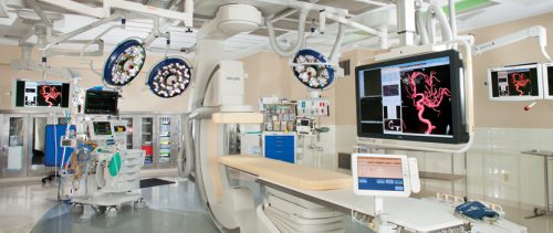
What is this, you ask? It’s a mashup of an operating room and an interventional radiology suite. It’s new. It’s big. It’s cool (literally, which is an issue for trauma surgeons).

More and more hospitals are adding hybrid rooms at the request of their vascular surgery teams. These rooms allow for both angiographic and open operative procedures, potentially at the same time. They are perfect for endovascular procedures that need some degree of hands-in work as well. They are frequently used for thoracic endovascular repair of the aorta (TEVAR), repair of abdominal aortic aneurysm (AAA), and transcatheter aortic valve replacement (TAVR).

These rooms would seem to be perfect for some trauma cases as well. Some injuries require a mix of interventional work and open surgery. Think complex pelvic fractures and extremity vascular injuries.

But before you go rushing off to the hybrid room with the next patient you think might benefit from it, consider these issues:

  • You must first secure access to the hybrid room. Just because you want it doesn’t mean you can get it. This room was probably built with other services in mind. You must work with them closely to set up rules and priorities. Consider questions like, can a trauma case bump an elective one?
  • Decide what specific cases can be done in the room. Don’t waste it on procedures that can be done in any old OR. Ideally, it is for multi-team cases and must take advantage of the radiographic capabilities of the hybrid room. If it doesn’t, it should be done in any other room of appropriate size.
  • Check your hardware. Make sure that anything you might attach to the hybrid table actually will attach to it. Frequently, the side rails are missing and the table thickness is different than a standard OR table. Check all of your retractor systems for compatibility. If your neurosurgeons use a skull clamp like a Mayfield, make sure it will attach to the table. If they do not, look for adapters to make it possible. Don’t discover this on your first trip to the room.
  • Watch for hypothermia! These are big rooms, and are difficult to heat up uniformly. In addition, the electronics in the room may be heat sensitive, so you may not be able to raise the temperature to the levels you are accustomed. Place heating systems under and around the patient as much as possible, warm everything that goes into them, and monitor their temp closely.
  • Treat the equipment with respect.  This stuff is delicate, and must be used by other surgeons for sensitive procedures. Don’t break it!

Related posts: