Category Archives: Head

Do We Really Need To Consult Neurosurgery For Mild TBI?

We consult our neurosurgeons too often. Think back on all the head injured patients you have admitted and placed a neurosurgical consult. How many times did they recommend something new or different, or take them to surgery? Not very often, I would guess.

This is becoming a hot topic. Check out the references below to read about a few other studies that have taken a similar approach.

The trauma group at Scripps Mercy in San Diego retrospectively reviewed their admissions to determine how often patients with mild TBI (GCS > 13) and some degree  intracranial hemorrhage required neurosurgical intervention, even if they were intoxicated or taking anti-platelet or anticoagulant drugs. A total of 500 patients were studied over a 28 month period.

Here are the factoids:

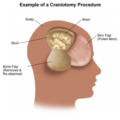
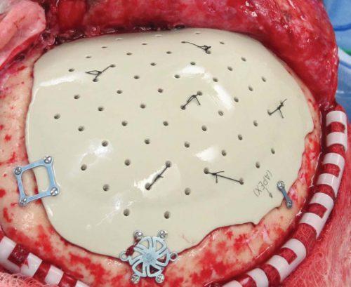
  • 49 (10%) of patients required some sort of neurosurgical intervention (41 craniotomy/craniectomy, 8 ICP monitors)
  • 93% of patients had neurosurgical consultation, and made additional recommendations in only 10 (2%),none of which changed management
  • There was no clinical difference in GCS between those who received an intervention and those who did not
  • Epidural and subdural hematomas were significant predictors of neurosurgical intervention
  • Intoxication or use of anti-platelet or anticoagulant drugs was not associated with intervention. These were present in 30% of all patients!
  • Unsurprisingly, ICU and hospital length of stay were longer in patient who underwent an intervention

Bottom line: As I said, this seems to be a hot research topic. And in this study, the numbers are getting larger and the criteria more inclusive (alcohol and anticoagulants allowed).

Neurosurgeons play a very important role in patients with more moderate to injury to their brain, and with spine injuries. But their input may not be needed in many patients with milder injuries. These data suggest that, in patients with GCS > 13, only subdural and epidural hematomas require consultation because they are much more likely to require intervention. 

This parallels a practice guideline we have in place where patients with subarachnoid or small intraparenchymal hemorrhage, or a linear skull fracture are managed by the trauma service without neurosurgical consultation. We do involve them if there is any intracranial hemorrhage with a history of anticoagulant use, however.

We all need to use our neurosurgeons wisely, and this paper helps to clarify situations where they may and may not be needed. 

Related posts:

Reference: Routine neurosurgical consultation is not necessary in mild blunt traumatic brain injury. J Trauma 82(4):776-780, 2017

Everything You Wanted To Know About: Cranial Bone Flaps

Patients with severe TBI frequently undergo surgical procedures to remove clot or decompress the brain. Most of the time, they undergo a craniotomy, in which a bone flap is raised temporarily and then replaced at the end of the procedure.

But in decompressive surgery, the bone flap cannot be replaced because doing so may increase intracranial pressure. What to do with it?

There are four options:

  1. The piece of bone can buried in the subcutaneous tissue of the abdominal wall. The advantage is that it can’t get lost. Cosmetically, it looks odd, but so does having a bone flap missing from the side of your head. And this technique can’t be used as easily if the patient has had prior abdominal surgery.

2. Some centers have buried the flap in the subgaleal tissues of the scalp on the opposite side of the skull. The few papers on this technique demonstrated a low infection rate. The advantage is that only one surgical field is necessary at the time the flap is replaced. However, the cosmetic disadvantage before the flap is replaced is much more pronounced.

3. Most commonly, the flap is frozen and “banked” for later replacement. There are reports of some mineral loss from the flap after replacement, and occasional infection. And occasionally the entire piece is misplaced. Another disadvantage is that if the patient moves away or presents to another hospital for flap replacement, the logistics of transferring a frozen piece of bone are very challenging.

4. Some centers just throw the bone flap away. This necessitates replacing it with some other material like metal or plastic. This tends to be more complicated and expensive, since the replacement needs to be sculpted to fit the existing gap.

So which flap management technique is best? Unfortunately, we don’t know yet, and probably never will. Your neurosurgeons will have their favorite technique, and that will ultimately be the option of choice.

Reference: Bone flap management in neurosurgery. Rev Neuroscience 17(2):133-137, 2009.

Rest vs Physical Activity After Mild Pediatric Concussion: Which Is Better?

One of the most common recommendations after a child or young adult sustains a mild TBI is to rest. And even better, brain rest. I’ve written about that topic several times over the years.

But what about physical rest? There is a large body of literature documenting the numerous mental and physical benefits of exercise. Couldn’t they also apply after concussive injury to the brain? A study published recently tried to determine if physical activity or lack of it after mild TBI was helpful in reducing the incidence of post-concussive symptoms.

This was a planned analysis of prospectively collected data from nine research network hospital emergency departments in Canada. Children from age 5 through 17 were enrolled if they had received a concussion within 48 hours of the ED visit, as defined by the 2012 Zurich consensus. They were excluded if they had a positive head CT, GCS < 14, or pre-existing cognitive deficits.

Initial research data was collected during the ED visit, and followup phone calls were made by the research team at 7 and 28 days. They asked about self-reported level of physical activity on day 7, and post-concussive symptoms and their change over time on days 7 and 28.

Here are the factoids:

  • Of 3063 patients enrolled, 84% completed the ED assessment. 171 were excluded because they could not be contacted for the activity assessment on day 7.
  • Post-concussive symptoms were present in 30% of these children overall
  • 70% participated in physical activity during the first week: 32% light aerobic, 9% sport-specific, 6% non-contact drills, 4% full-contact practice, and 18% full competition (ignoring doctor’s orders?)
  • Overall, early activity was associated with a lower risk of post-concussive symptoms (25% vs 44%)
  • In patients who were symptomatic at day 7, symptoms were decreased at 28 days in patients who engaged in light aerobic activity, moderate activity, and even full-contact activity

Bottom line: This was a well designed study, but obviously with a number of limitations. Physical activity was self-reported, there may have been other factors that could not be controlled, and the study did not inquire about activity between days 7 and 28.

But this study appears to suggest that, like in most other areas, exercise is good. Even for the brain recovering from a concussion. Obviously, a really good randomized study would be the gold standard, but I doubt that will be done anytime soon. Trauma professionals may want to consider a cautious return to light to moderate activity as soon as the child feels well enough. But keep in mind that, in general, the onset of fatigue is a good indicator that it is time to stop activity and rest. And full contact should probably be avoided, especially because of the risk of re-injury.

Related posts:

Reference: Association between early participation in physical activity following acute concussion and persistent postconcussive symptoms in children and adolescents. JAMA 316(23):2504-2514, 2016.

EAST 2017 #8: When Is “Mild TBI” Not So Mild?

Traumatic brain injury (TBI) is very common, with the majority falling into the “mild” category. This is usually defined as patients with injury to the head and a GCS of 13-15. These uncomplicated patients are frequently discharged from the emergency department, or undergo only a brief evaluation if admitted for other reasons.

The group at Shock Trauma focused on a less appreciated subset of mild TBI patients, those whose condition is a little more complicated. Specifically, these are patients with GCS 13-14 with positive findings on head CT leading to a calculated abbreviated injury score (head) of > 2, and some persistence of their symptoms while in the hospital. At many hospitals (including my own), these patients receive an inpatient TBI evaluation. But if they pass this initial screening, they are not consistently referred for any outpatient TBI followup.

Are these mild, complicated TBI patients (mcTBI) unique? Do they behave the same as the uncomplicated ones? The research group performed a prospective study on patients who sustained an mcTBI over a 4 month period.  They excluded patients with mental illness, dementia, and non-English speaking and homeless patients. They tried to contact patients up to three times after discharge to administer several standard tests and determine if they had any specific residual symptoms.

Here are the factoids:

  • Of the 142 patients with mcTBI during the study period, there was substantial attrition over time, with only 25 remaining at 6 months and 10 at one year
  • 64% of patients who responded at 6 months remained symptomatic. Depression, dizziness, and a feeling of impaired health were common.
  • 80% of patients still described symptoms at one year. The same complaints were most common, and some required changes in activities of daily living or assistive devices.

Bottom line: Although small and fraught with the usual problems in long-term tracking of urban trauma patients, this study is eye-opening. We too often dismiss “mild TBI” and being almost nothing, even in patients with positive findings on head CT. This work suggests that we are underestimating the needs of those patients. The authors used this data to design longer-term care processes for this subset of patients. Other centers should follow suit to make sure these patients’ post-injury needs are better met.

Questions and comments for the authors/presenters:

  • Describe the possible biases that patient selection and attrition may have had on the study
  • What type of TBI screening do you use in the hospital?
  • Given that a number of assessments were administered over the phone, I look forward to hearing some of the other details not listed in the abstract
  • Was there any correlation between specific CT findings and later symptoms?
  • Provide details of your long-term care programs for these patients
  • I enjoyed this thought provoking abstract!

Click here to go the the EAST 2017 page to see comments on other abstracts.

Related posts:

Reference: Mild TBI is not ‘mild’… survivors tell their complicated stories. Quick Shot #3, EAST 2017.

EAST 2017 #5: Subarachnoid Hemorrhage, Neurosurgical Consults, and Repeat Head CT

Neurosurgical involvement in the management of simple traumatic brain injury (TBI) has been slowly dwindling over the past several years. This is the result of the general consensus that very few of these patients progress to need neurosurgical procedures.

A group at Wright State University in Dayton sought to define the progression of one specific finding in TBI, the subarachnoid hemorrhage (SAH). Secondarily, the wanted to determine if a neurosurgery consultation was warranted in these patients.

They performed a five year retrospective review of their registry data, identifying patients with both mild TBI (GCS 13-15) and SAH. They excluded patients with any other brain lesion on CT.

Here are the factoids:

  • 301 patients were enrolled during the 5 year period
  • All had a neurosurgical consultation
  • Time between the initial CT and a followup scan was about 11 hours
  • 91% showed stable or resolving SAH on the followup scan
  • 9% showed a worsening SAH or additional lesions on the repeat scan

Bottom line: The authors conclude that initial neurosurgical consultation is not needed, since only 9% of patients have worrisome findings on repeat CT. They do, however, recommend that the practice of repeat scanning be continued because of this same number.

Our trauma service looked at this issue a year ago, and determined that most of these lesions either do not progress, or never require any intervention if they do, with a few notable exceptions. For that reason, we abandoned both neurosurgical consultation and repeat CT scans for patients with non-aneurysmal SAH, a single parenchymal hemorrhage, or linear skull fractures. We continue to do both for patients with epidural and/or subdural hemorrhage. You can download a copy of this protocol here.

Questions and comments for the authors/presenters:

  • Did you look at platelet count or INR in the study. Were patients excluded based on abnormal values?
  • Did every patient get a repeat scan?
  • Break down the lesions in the 9% of patients who had some sort of progression or new finding. Did you see any common themes (age, chronic alcohol use, etc.)?
  • Did you encounter any patients with “non-central SAH”, that might indicate an aneurysm? How were they dealt with?
  • How has or will your trauma service change its practice based on your findings.

Click here to go the the EAST 2017 page to see comments on other abstracts.

Related posts:

Reference:   Management of subarachnoid hemorrhage (SAH) by the trauma service: are repeat CT scanning & routine neurosurgical consultation necessary? Poster #16, EAST 2017.