Category Archives: General

The Flat Vena Cava Sign? Published and Revisited

I’ve previously blogged about the flat vena cava sign as an indicator of low volume status in trauma patients. And I commented on this paper when it was presented at EAST, which had a surprisingly negative result. It’s now been vetted by peer reviewers and published, and I’ve had the opportunity to read through the entire manuscript (always important). So let’s take a second look now.

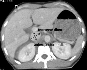
A retrospective study at George Washington University was carried out over a one year period. They looked at all of their highest level trauma activation patients who also underwent CT scan of the abdomen. Images were read by three radiologists and inter-rater reliability was reviewed. The transverse to anteroposterior diameter ratios were calculated to determine flatness.

Here are the factoids:

  • 276 patients met enrollment criteria, and were mostly male and blunt trauma
  • The IVC was nearly round in 21% of patients and collapsed in 26%
  • There was no association between IVC shape and shock index, blood pressure, Hbg, lactate, urgent operation, angiography or length of stay
  • There was also no association between IVC shape and blood transfusion or death
  • Correlation of the reads between radiologists was good

So what gives? A paper I reviewed three years ago in the Journal of Trauma came to a different conclusion. They found that a flat IVC on CT scan (defined as a transverse to AP ratio of 4:1 or greater) was associated with a significantly higher chance of receiving more crystalloid or blood, as well as requiring an operation within 24 hours. 

This newer paper was able to look at a larger group of patients, and they were able to tease out why it initially looked like the flat cava looked like a good predictor for bad things to come. The problem was statistical skewing from a few extreme outliers. When properly corrected, it completely changed things. And looking at the older study, it appears that outliers may have also been the reason for the positive result. This is why I encourage everyone to always read the entire paper! The older paper involved a smaller series (114 patients), but it was prospective and seemed to have reasonable statistical analyses.

image

Bottom line: It looks like the flat vena cava sign, as measured by a static CT, should be discarded as an indicator of impending shock. Whether or not a more dynamic look (using ultrasound) is valuable remains to be determined.

Related post:

Reference: Inferior vena cava size is not associated with shock following injury. J Trauma 77(1):34-39, 2014.

Print Friendly, PDF & Email

Phlebotomy And Pediatric Solid Organ Injury

I recently wrote about this journal article from a couple of pediatric trauma programs in New York. The article tried to focus on reducing the rate of phlebotomy in children who are being observed for solid organ injury. I was more excited about the overall protocol being used to manage liver and spleen injury, as it was a great advance over the original APSA guideline. But let’s look at the phlebotomy part as well.

This is an interestingly weird study, and you’ll see what I mean shortly. Two New York trauma hospitals that take care of pediatric patients pooled 4 years of registry records on children with isolated blunt liver and/or spleen injuries. Then they did a tabletop excercise, looking at “what if” they had applied the APSA guideline, and “what if” they had applied their new, proposed guideline.

Interestingly, this implies that they were using neither! I presume they are trying to justify (and push all their partners) to move to the new protocol from (probably) random, individual choice.

Here are the factoids:

  • 120 records were identified across the 2 hospitals that met criteria
  • Late presentation to the hospital, contrast extravasation, comorbidities, lack of imaging, operative intervention at an outside hospital excluded 59 patients, leaving 61 for analysis. Three of those patients became unstable and were also excluded.
  • None of the remaining patients required operation or angioembolization
  • Use of the “new” (proposed) protocol would reduce ICU admissions by 65%, reduce blood draws by 70%, and reduce hospital stay by 37%
  • Conclusion: use of the protocol would eliminate the need for serial phlebotomy (huh?)

Bottom line: Huh? All this to justify decreasing blood draws? I know, kids hate needles, but the data on decreased length of stay in the hospital and ICU is much more important! We’ve been using a protocol similar to their “new” one at Regions Hospital, which I’ve shared below. We’ve been enjoying decreased resource utilization, blood draws, and very short lengths of stay for over a decade. And our analysis showed that we save $1000 for every patient entering the protocol, compared to the old-fashioned and inefficient way we used to manage them.

Related posts:

Reference: Reducing scheduled phlebotomy in stable pediatric patients with liver or spleen injury. J Ped Surg 49(5):759-762, 2014.

Print Friendly, PDF & Email

Pediatric Solid Organ Injury Management: It’s About Time!

There was an interesting article released in the Journal of Pediatric Surgery in May about spleen and liver injury management in children. It’s interesting because if you just look at the title, you might just skip over it. The title suggests that it describes reducing scheduled phlebotomy in kids who are undergoing solid organ injury management. But the real meat of this article has to do with the protocol they are using to treat the children.

Nonoperative management of these injuries in children started becoming popular 40 years ago (!). But for decades, everyone put their own spin on how to do it. Bed rest for a week (or more). NPO for days! Limited physical activity for extended periods. Then the American Pediatric Surgery Association (APSA) published a set of guidelines about 15 years ago that took some of the guesswork out of it.

Although nonoperative management of these injuries in kids preceded its adoption in adults by a nearly two decades, it has languished in the APSA format for quite some time. Many pediatric surgeons still use these guidelines, even though adult spleen and liver injury management have advanced to shorter and more streamlined care.

We adopted a solid organ management guideline at Regions Hospital over ten years ago, and have made a few minor tweaks over the years. Nowadays, our grade I-III injuries can be home as early as 36 hours after admission, and frequently are. Grades IV and V are eligible to be discharged after just 24 more hours if they have no other injuries to keep them in the hospital.There are very rare failures.

I’ll detail the factoids about the phlebotomy part of this paper in tomorrow’s post. But I do want to show you the more aggressive protocol the authors are using (one of whom authored the original APSA guideline).

Here it is:

image

Bottom line: Note how quickly children are allowed to get up, eat, and get out of the hospital using the “new” protocol. Many adult centers have been using similar ones for years. It’s nice to see that adult and pediatric protocols are finally beginning to converge. After all, we figured out our current adult management based on our experience with kids 30 years ago! 

Related posts:

Reference: Reducing scheduled phlebotomy in stable pediatric patients with liver or spleen injury. J Ped Surg 49(5):759-762, 2014.

Print Friendly, PDF & Email

Another Way To Treat Tension Pneumothorax

Kenji Inaba and colleagues have done a lot of work on tension pneumothorax (tPTX) in the past few years. They’ve looked for the best devices and the best positions on the chest to quickly and effectively treat this emergency. Now, they’ve published a study on using what looks like a “better mousetrap” for relieving tension physiology.

Previous work from this lab has shown that up to a quarter of needle thoracostomies fail within 5 minutes due to mechanical reasons. This leaves a small window for insertion of the real chest tube. And even though much of the pressure may be relieved, a significant amount of air may be left in the chest, impeding recovery from PEA arrest.

They looked at the use of a 5mm laparoscopy port for relief of tension pneumothorax in Yorkshire swine. The exact size of the pigs was not listed, but these animals weigh 25 pounds at 6 weeks of age, and the pictures in the article show a reasonable sized animal. I’m not sure they were 70kg, though.

Here are the factoids:

  • Five animals were used, and 30 episodes of tPTX and 27 episodes of PEA arrest from tPTX
  • Tension pneumothorax was created by insufflating the chest with CO2 using a 10mm laparoscopic trocar
  • tPTX was completely relieved by insertion of the 5mm trocar in 100% of trials, with all physiologic measures returning to baseline within 1 minute
  • Circulation was restored to normal within 30 seconds in 100% of trials
  • There was no damage to heart or lung from trocar placement in any of the 5 animals

image

Bottom line: Once again, Inaba and crew have added some interesting tidbits to our knowledge base. You already know I’m not a fan of animal studies like this, but this one lays the ground work for some work in humans. We still need to know how the “usual American body habitus” will affect the use of this device. The only downside is the expense of the trocar, which is a lot more than a simple long needle. But if it is as efficacious in humans as it is in pigs, it may be worth it!

Related posts:

Reference: Standard laparoscopic trocars for the treatment of tension pneumothorax: A superior alternative to needle decompression. J Trauma 77(1):170-175, 2014.

Print Friendly, PDF & Email

Procedural Complications: Residents vs Advanced Practice Providers

With the implementation of resident work hour restrictions 10 years ago, resident participation in clinical care has declined. In order to make up for this loss of clinical manpower and expertise, many hospitals have added advanced clinical providers (ACPs, nurse practitioners and physician assistants). These ACPs are being given more and more advanced responsibilities, in all clinical settings. This includes performing invasive procedures on critically ill patients. 

A recent study from Carolinas Medical Center in Charlotte NC compared complication rates for invasive procedures performed by ACPs vs residents in a Level I trauma center setting.

A one year retrospective study was carried out. Here are the factoids:

  • Residents were either surgery or emergency medicine PGY2s
  • ACPs and residents underwent an orientation and animal- or simulation-based training in procedures
  • All procedures were supervised by an attending physician
  • Arterial lines, central venous lines, chest tubes, percutaneous endoscopic gastrostomy, tracheostomy, and broncho-alveolar lavage performances were studied
  • Residents performed 1020 procedures and had 21 complications (2%)
  • ACPs performed 555 procedures and had 11 complications (2%)
  • ICU and hospital length of stay, and mortality rates were no different between the groups

Bottom line: Resident and ACP performance of invasive procedures is comparable. As residents become less available for these procedures, ACPs can (and will) be hired to  take their place. Although this is great news for hospitals that need manpower to assist their surgeons and emergency physicians, it should be another wakeup call for training programs and educators to show that resident education will continue to degrade.

Related posts:

Reference: Comparison of procedural complications between resident physicians and advanced clinical providers. J Trauma 77(1):143-147, 2014.

Print Friendly, PDF & Email