Category Archives: General

Gunshots And CT Scan Of The Abdomen

Abdominal gunshots and CT scanning are usually thought to be mutually exclusive. The usual algorithm generally means a prompt trip to the operating room. But as with many things in the management of trauma, there are always exceptions. The key is to understand when exactly one of those exceptions is warranted.

Exception 1: Did it really enter the abdomen? Gunshots have enough energy that they usually do get inside. However, freaky combinations of trajectory and body habitus do occur. There are three tests that must be passed in order to entertain the possibility that the bullet may not have made it inside your patient: physiology, anatomy, and physical exam. For physiology, the patient must be completely hemodynamically stable. Anatomically, the trajectory must make sense. If the known wounds and angles allow a tangential course make sense, then fine. But if there is a hole in the epigastrium and another next to the spine, you have to assume the bullet went straight through. Finally, the physical exam must be normal. No peritonitis. No generalized guarding. Focal tenderness only in the immediate area of any wounds. If all three of these criteria are passed, then a CT can be obtained to demonstrate the trajectory.

Exception 2: Did it enter an unimportant area of the abdomen? Well, there’s really only one of these, and that’s the area involving the right lobe of the liver and extending posteriorly and lateral to it. If the bullet hole(s) involve only this area, and the three tests above are passed, CT may confirm an injury that can be observed. However, there should only be a minimal amount of free fluid, and no soft tissue changes of any kind adjacent to bowel.

Exception 3: A prompt trauma lap was performed, but you think you need more information afterwards. This is rare. The usual belief is that the eyes of the surgeon provide the gold standard evaluation during a trauma lap. For most low velocity injuries with an easily understood trajectory, this is probably true. However, high velocity injuries, those involving multiple projectiles, or complicated trajectories (side to side) can be challenging for even the most experienced surgeon. Some areas (think retroperitoneum or deep in the pelvis) are tough to visualize completely, especially when there’s blood everywhere. These are also the cases most likely to require damage control surgery, so once the patient has been temporarily closed, warmed and resuscitated, a quick trip to CT may be helful in revealing unexpected shrapnel, unsuspected injuries, or other issues that may change your management. Even a completely unsurprising scan can provide a higher sense of security.

Bottom line: CT of the abdomen and gunshots to that area may actually coexist in some special cases. Make sure the physiology, anatomy and physical exam criteria are passed first. I also make a point of announcing to all trainees that taking these patients to CT is not the norm, and carefully explain the rationale. Finally, apply the concept of the null hypothesis to this situation. Your null hypothesis should state that your patient does not need a CT after gunshot to the abdomen, and you have to work to prove otherwise!

How To: Secure An Endotracheal Tube To… Nothing!

Several decades ago I took care of a patient who posed an interesting challenge. He had been involved in an industrial explosion and had sustained severe trauma to his face. Although he was able to speak and breathe, he had a moderate amount of bleeding and was having some trouble keeping his airway clear.

Everyone frets about getting an airway in patients who have severe facial trauma. However, I find it’s usually easier because the bones and soft tissue move out of your way. That is, as long as you can keep ahead of the bleeding to see your landmarks.

In this case, the intubation was easy. The epiglottis was visible while standing above the patient’s head, so a laryngoscope was practically unnecessary! But now, how do we secure the tube so it won’t fall out? Sure, there are tube-tamer type securing devices available, but what if they are not available to you? Or this happened in the field? Or it was in the 1980’s and it hadn’t been invented, like this case?

The answer is, create your own “skin” to secure the tube to. Take a Kerlix-type stretchable gauze roll and wrap it tightly around their head. Remember, they are sedated already and they can breathe through the tube. This also serves to further slow any bleeding from soft tissue. Once you have “mummified” the head with the gauze roll, tape the tube in place like you normally would, using the surface of the gauze as the “skin.”

Be generous with the tape, because the tube is your patient’s life-line. Now it’s time for the surgeons to surgically stabilize this airway, usually by converting to a tracheostomy.

Related posts:

Gunshot To The Face!

You’ve just been pre-notified of an incoming trauma activation: gunshot to the face. No other information. How concerned should you be? Here are some things to think about as you wait for the patient to arrive:

  • Is it really a gunshot? Sometimes shotgun injuries are reported as gunshots. Big difference!
  • Will I need to preserve evidence? In general, yes. In most cases other than suicide attempts, there is probably a good chance that criminal activity was involved. Be prepared to preserve all patient belongings in paper bags, and have a chain of custody form available.
  • Am I and my team safe? There is a possibility that someone wants your incoming patient dead. They may want to finish the job, in you emergency department. Make sure the area is secure.

Once the patient arrives, it’s best to think through things via the ATLS framework.

  • Airway. If the injury involves the lower part of the face or neck, make sure the airway is safe and/or secure. Blood may create problems, as can edema from injury to soft tissues, especially in the floor of the mouth.
  • Breathing. Not a problem with these injuries unless significant aspiration has occurred. 
  • Circulation. The face can really bleed, and only a few areas are amenable to the usual surgical control (clamping, tying). Direct pressure must be used for the rest, and this doesn’t always work. Bleeding from sinuses may be controlled with packing or the foley catheter trick (inserted through bullet tract). But if you can’t stop it, then it’s time to expedite to the OR.
  • Disability. You do have to worry about the cervical spine if the path of the bullet is not obvious. If the patient is stable, immobilize the neck and use the CT scanner to see if any fragments involved the spine. If you must run to the OR with an unstable patient, then try to quickly shoot an old-fashioned cross-table lateral. This will give you quick and dirty info on how much you can manipulate the neck.

Related posts:

Pneumomediastinum After Falling Down

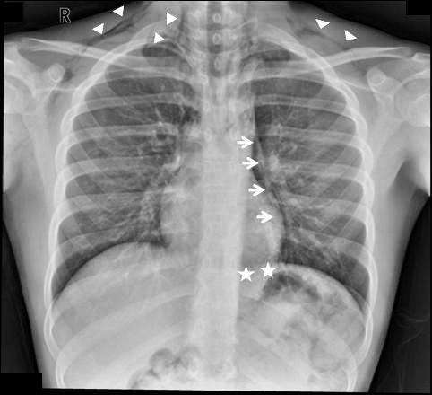
Finding pneumomediastinum on a chest xray or CT scan always gets one’s attention. However, seeing this condition after a simple fall from standing is very simple to evaluate and manage.

There are 3 potential sources of gas in the mediastinum after trauma:

  • Esophagus
  • Trachea
  • Smaller airways / lung parenchyma

Blunt injury to the esophagus is extremely rare, and probably nonexistent after just falling down. Likewise, a tracheal injury from falling over is unheard of. Both of these injuries are far more common with penetrating trauma.

This leaves the lung and smaller airways within it to consider. They are, by far, the most common sources of pneumomediastinum. The most common pattern is that this injury causes a small pneumothorax, which dissects into the mediastinum over time. On occasion, the leak tracks along the visceral pleura and moves directly to the mediastinum.

Management is simple: a repeat chest xray after 6 hours is needed to show non-progression of any pneumothorax, occult or obvious. This image will usually show that the mediastinal air is diminishing as well. There is no need for the patient to be kept NPO or in bed. Monitor any subjective complaints and if all progresses as expected, they can be discharged after a very brief stay.

January Trauma MedEd Newsletter Released

The January newsletter is now available! Click the image below or the link at the bottom to download. This month’s topic is “Shock”.

In this issue you’ll find articles on:

  • Thoughts on trauma patient stability
  • Does initial hematocrit predict shock?
  • Not all plasma is created equal
  • Can I take a hypotensive patient to CT?
  • Pelvic fractures: OR vs angio in the unstable patient

Subscribers received the newsletter first at the end of last week. If you want to subscribe (and download back issues), click here.

Click here to download the current issue.