Category Archives: General

Extraperitoneal Bladder Rupture

This injury is likely to occur in patients who have a full bladder and sustain anterior pelvic trauma that typically leads to fractures. They generally present with gross hematuria upon placement of the bladder catheter. This should prompt an abdominal CT scan with cystogram technique.

CT cystogram involves pressurizing the bladder with contrast prior to the study. This differs from the usual method of clamping the catheter and allowing the bladder to passively fill. The literature here is clear: failure to use cysto technique will miss 50% of these injuries.

The majority of extraperitoneal bladder injuries can be treated nonoperatively, and probably do not need Urology involvement. The bladder catheter is left in place 10-14 days (we do 10 days), and a repeat cystogram is obtained. If there is no leak, the catheter can be removed. If there is still some leakage, Urology consultation should then be obtained. 

There are a few cases where operative management is required:

  • There is some intraperitoneal component of bladder injury
  • Fixation of the pubic rami is required (bathing the orthopedic hardware with urine is frowned upon)
  • Failure of conservative management

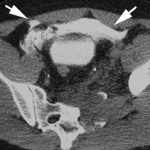
Arrows in the photo show extraperitoneal extravasation of cystogram contrast.

Related posts:

Warm Water Immersion Policy for Hypothermia

A few days ago, I wrote about using a therapy tank for immersion to rapidly rewarm patients (click here to read it). Since this type of management usually means moving out of the ED to a separate patient care are, it is important to have a policy that spells out responsibilities for all personnel involved. 

Click here or click the image above to download a copy of the Regions Hospital Trauma Program policy.

Trauma 20 Years Ago: CT Imaging of the Aorta

CT scan is now the standard screening test for injury to the thoracic aorta. But 20 years ago, we were still gnashing our teeth about how to detect this injury.

An interesting paper was published in the Journal of Trauma 20 years ago this month on this topic. Over a 2 year period, the Medical College of Wisconsin at Milwaukee looked at all patients who underwent imaging for aortic injury. At the time the gold standard was aortogram. They looked at patients who underwent this study and CT, which was not very common at the time.

They had 50 patients who underwent aortography alone and 17 who underwent both tests. Of the 17, 5 had the injury, but only three were seen on CT. There were also two false positives. Sensitivity was 83%, specificity was 23%, with 53% accuracy. The authors concluded that any patients with strong clinical suspicion of aortic injury should proceed directly to aortogram.

Why the difference today? Scan technology and resolution has increased immensely. Also, the timing of IV contrast administration has been refined so that even subtle intimal injuries can be detected. CT scan is now so good that we have progressed from the CV surgeon requiring an aortogram before they would even consider going to the OR, to the vascular surgeon / interventional radiologist proceeding directly to the interventional suite for endograft insertion.

Solid Organ Injury: How Soon Can We Begin Chemical DVT Prophylaxis?

Nonoperative management of solid organ injury is the norm, and has reduced the operative rate significantly. At the same time, the recognition that development of deep venous thrombosis (DVT) in trauma patients is commonplace creates uncertainty? Is it safe to give chemical prophylaxis with low molecular weight heparin (LMWH)? How soon after injury?

The trauma group at USC+LAC recently published the findings of a retrospective review of 312 patients undergoing nonoperative management for their liver, spleen or kidney injuries. They looked at chemical prophylaxis administration and its relationship to failure of nonop management of solid organ injury.

As expected, as the grade of the solid organ injury increased, so did the failure rate of nonoperative management. Administration of low molecular weight heparin, such as enoxaparin, did not increase failure rate in this study. All but one failure occurred in patients who had not yet received the injections. Likewise, two DVT and two pulmonary embolisms occurred, but only in patients who had not yet received prophylaxis. 

Bottom line: This small study offers some assurance that early prophylaxis is okay, and a few prospective studies do exist. UCSF / San Francisco General is comfortable beginning chemical prophylaxis 36 hours postop, regardless of solid organ injury. Look for more guidance on this issue in the coming year or so. Until then, consider starting LMWH prophylaxis early to avoid complications from DVT or PE.

Reference: Thromboembolic prophylaxis with low-molecular-weight heparin in patients with blunt solid abdominal organ injuries undergoing nonoperative management: current practice and outcomes. J Trauma 70(1): 141-147, 2011.