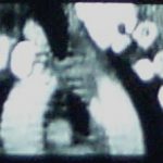
Trauma 20 Years Ago: CT Imaging of the Aorta
CT scan is now the standard screening test for injury to the thoracic aorta. But 20 years ago, we were still gnashing our teeth about how to detect this injury.
An interesting paper was published in the Journal of Trauma 20 years ago this month on this topic. Over a 2 year period, the Medical College of Wisconsin at Milwaukee looked at all patients who underwent imaging for aortic injury. At the time the gold standard was aortogram. They looked at patients who underwent this study and CT, which was not very common at the time.
They had 50 patients who underwent aortography alone and 17 who underwent both tests. Of the 17, 5 had the injury, but only three were seen on CT. There were also two false positives. Sensitivity was 83%, specificity was 23%, with 53% accuracy. The authors concluded that any patients with strong clinical suspicion of aortic injury should proceed directly to aortogram.
Why the difference today? Scan technology and resolution has increased immensely. Also, the timing of IV contrast administration has been refined so that even subtle intimal injuries can be detected. CT scan is now so good that we have progressed from the CV surgeon requiring an aortogram before they would even consider going to the OR, to the vascular surgeon / interventional radiologist proceeding directly to the interventional suite for endograft insertion.


