Category Archives: General

Delayed Splenic Rupture: Part 2

Yesterday I wrote about the history of “delayed splenic rupture.” Today I’ll discuss how to deal with it.

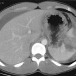
If possible, try to avoid ever having to mess around with this clinical problem. If you order an abdominal CT after blunt trauma and see a splenic contrast blush of either type (pseudoaneurysm or extravasation, see left photo), then deal with it before the patient even knows he has a problem. A trip to interventional radiology will usually solve the problem. And if embolized, these patients almost never come back with a bleeding problem.

As I’ve said many times before, if the patient is hemodynamically compromised, then an OR visit is required. The usual solution is splenectomy. Some recommend repairing the spleen, but this is technically more difficult than it sounds, and it is difficult for the surgeon to sleep soundly after performing one of these.

Lets say you inherited one of these from someone else, or ignored the warning signs on the initial CT. The usual time frame for presentation to the ED with acute bleeding is 7 to 10 days after the initial injury. If they are not stable, physical exam or FAST will quickly direct you to the OR, once again for splenectomy. Some patients will stabilize with fluids and can safely be sent to CT scan.

Once the CT confirms what the problem is, a trip to interventional radiology is in order if the patient remains stable. Here is the key: the radiologist must embolize something! If they find a bleeding vessel, then they can selectively embolize it. If they don’t, then the main splenic artery should be embolized. This will decrease the arterial pressure head, but won’t eliminate it. It will decrease the likelihood of additional bleeding as much as possible.

At this point, the patient should be admitted to the trauma service and monitored using your solid organ injury protocol. If they have any hemodynamic issues, it’s time to remove the spleen. Remember, this is the third time they’ve had a problem, and like in baseball, their spleen is out! Attempted splenorrhaphy at this point is pointless and may lead to yet another operation.

Related posts:

Print Friendly, PDF & Email

Delayed Splenic Rupture: Part 1

This post was prompted by a paper that somehow got into the Journal of Trauma this month on nonoperative management of delayed splenic rupture after trauma. It’s a bad retrospective review of 15 patients which I’ll say more about tomorrow. There’s very little good literature on this topic, so I wanted to share some personal observations.

Back in the days before CT scan (and unfortunately, I remember them), the diagnosis of abdominal injury was much more difficult. It was primarily qualitative, meaning that we somehow figured out that they either had it or they didn’t. We could not very easily figure out what specific injuries a given patient had. However, management was simple: we went to the operating room, found out and fixed it.

Sometimes, though, we would encounter a patient who had been involved in some type of blunt trauma a week or two earlier who presented to the ED with left-sided abdominal pain, shock and anemia. The diagnosis was “delayed splenic rupture” and they were taken to OR for a splenectomy.

When CT scan came along, we found out that these were actually “delayed recognition of splenic injury.” We still took them to the OR for splenectomy, but with experience this slowly gave way to splenic repair, and then to nonoperative management. 

There is still one subset of these injuries that is problematic: spleen injury with a contrast blush. It turns out that there are really two types of blush: contrast seen within a pseudoaneurysm within the splenic pulp, and extravasation. And furthermore, the pseudoaneurysm is the culprit in most “delayed splenic ruptures.”

Tomorrow, I’ll write about how to recognize this potential problem, what to do about it acutely, and what to do if it was missed and the patient presents to your ED ten days later in shock.

Related post:

Reference: Nonsurgical management of delayed splenic rupture after blunt trauma. J Trauma 72(4):1019-1023, 2012.

Print Friendly, PDF & Email

New Technology: Help Brain Injured Patients To Talk

It is can be extremely difficult to communicate with some brain injured patients. Many have global damage that precludes the processing necessary to formulate thoughts. However, some may be able to think but can’t effectively make themselves understood. Patients with the “locked in” syndrome are a perfect example.

A company called NeuroVigil has developed technology and data analysis techniques for extracting a wealth of information from a single-channel EEG. The iBrain system uses two sensors that do not require being stuck to the head with adhesive. A simple elastic band can hold them in place. 

Last year, the company fitted the device on Stephen Hawking to begin testing and training the system to assist with his communication efforts. Currently, Hawking uses an IR sensor that detects twitches in his cheek. These are painstakingly translated into letters and then words that are spoken by a computer. The iBrain system is being trained to recognize words via his EEG patterns and should speed up his communication with the outside world.

If this technology pans out, it may be used to communicate with moderate to severely injured TBI patients who have expressive language problems. It could also be used to test for and communicate with patients who are “locked in.”

The video was recorded at TEDMED 2009. Much of the key information is presented beginning at 10:10 into the video.

I have no financial interest in NeuroVigil

Print Friendly, PDF & Email

New Technology: The End Of Handwashing?

All healthcare professionals are notoriously bad about washing their hands, especially doctors. A variety of things have been developed to help us keep our hands clean, including simple soap and water, barriers like gloves, and various gels and foams (which I swear I can taste in my mouth 10 minutes later, even though I’m pretty sure I’m not putting my fingers there).

A recently published paper from China is shining new light on this topic (get it?). Researchers developed a hand-held, battery-powered plasma flashlight that gets rid of bacteria on skin in a flash. It costs less than $100 to produce and runs on a 12V battery. 

This device was found to inactivate all bacteria in a 17-layer biofilm containing a very hardy organism, enterococcus faecalis. It does not produce UV radiation, and the exact mechanism for the bacterocidal effect is unclear. There was no adverse effect on skin.

The main drawbacks to this device are that it only produces a small area of plasma, and it takes 5 minutes to kill all the bacteria. But take this to the next logical step. Many of you are familiar with the Dyson Airblade hand dryers found at many airports. Suppose you could produce a more intense plasma field using a more robust power supply (power line or ambulance power system) in a device that you could just pass your hands through to disinfect them? 

And if you really want to improve compliance, hook the unit to the door control so the doctor can’t even walk into a patient room without passing his hands through it!

Reference: Inactivation of a 25.5µm Enterococcus faecalis biofilm by a room-temperature, battery-operated, handheld air plasma jet. Journal of Physics D: Applied Physics 45(2012):165205 (5p), 2012.

Print Friendly, PDF & Email

Best Of: Flying After Pneumothorax

Patients who have sustained a traumatic pneumothorax occasionally ask how soon they can fly in an airplane after they are discharged. What’s the right answer?

The basic problem has to do with Boyle’s Law (remember that from high school?). The volume of a gas varies inversely with the barometric pressure. So the lower the pressure, the larger a volume of gas becomes. Most of us hang out pretty close to sea level, so this is not an issue.

However, flying in a commercial airliner is different. Even though the aircraft may cruise at 30,000+ feet, the inside of the cabin remains considerably lower though not at sea level. Typically, the cabin altitude goes up to about 8,000 to 9,000 feet. Using Boyle’s law, any volume of gas (say, a pneumothorax in your chest), will increase by about a third on a commercial flight. 

The physiologic effect of this increase depends upon the patient. If they are young and fit, they may never know anything is happening. But if they are elderly and/or have a limited pulmonary reserve, it may compromise enough lung function to make them symptomatic.

Commercial guidelines for travel after pneumothorax range from 2-6 weeks. The Aerospace Medical Association published guidelines that state that 2-3 weeks is acceptable. The Orlando Regional Medical Center reviewed the literature and devised a practice guideline that has a single Level 2 recommendation that commercial air travel is safe 2 weeks after resolution of the pneumothorax, and that a chest xray should be obtained immediately prior to travel to confirm resolution.

Bottom line: Patients can safely travel on commercial aircraft 2 weeks after resolution of pneumothorax. Ideally, a chest xray should be obtained shortly before travel to confirm that it is gone. Helicopter travel is okay at any time, since they typically fly at 1,500 feet or less.

References:

  • Practice Guideline, Orlando Regional Medical Center. Air travel following traumatic pneumothorax. October 2009.
  • Medical Guidelines for Airline Travel, 2nd edition. Aerospace Medical Association. Aviation, Space, and Environmental Medicine 74(5) Section II Supplement, May 2003.
Print Friendly, PDF & Email