Category Archives: General

Trauma In Pregnancy 1: Introduction

Trauma in the pregnant patient is scary, but thankfully not very common. About 1 in 15 pregnant women sustain some type of significant injury. About half are from car crashes (most commonly if unrestrained), and 25% each are from assault (frequently domestic) and falls. Unfortunately, trauma is the leading cause of non-obstetric fetal death.

What makes this type of trauma problematic is two-fold, literally. First, you have two patients. And second, neither one has “normal” physiology. The mother has made numerous adaptations to accommodate the pregnancy, and the fetus is essentially a small parasite, totally dependent on her.

Let’s look at the impact of some of the more important maternal adaptations:

  • Total blood volume increases by about one liter. This would seem to be good, but since it is mainly dilutional with no real increase in total RBCs, and this is coupled with a lower mean blood pressure. Trauma professionals can easily underestimate blood loss.
  • Going into the third trimester, the uterus can compress the IVC when the mother is lying supine. but it is quite frequently forgotten. One of the first maneuvers when you suspect an advanced pregnancy is to bump the patient to her left. Do this even if the blood pressure is normal.
  • The pelvic veins are huge. Disruption from pelvic fractures or penetrating injury can lead to massive bleeding.
  • The stomach is probably full, and under pressure from below. This increases aspiration risk in women who have decreased mental status or need intubation.

And don’t forget the fetus:

  • The fetal/placental/uterine complex is one large, non-compressible unit with multiple shear planes within it. Blunt force will stress those planes, and may result in disruption of the uterus from pelvic veins (massive bleeding), or separation of the placenta from the uterus (abruption).
  • The fetus is totally dependent on the mother for survival, but the placenta will protect the mother first, shutting down fetal circulation if she becomes hypotensive or hypoxic.
  • The baby was designed to come into this world at full term. We have developed the technology to sustain life in smaller and more premature babies. The magic number of weeks keeps slowly decreasing, but preemie survival without complications is a challenge.

Tomorrow, we’ll move on and get to the fun stuff, predicting outcome after trauma in pregnancy.

Print Friendly, PDF & Email

Trauma Activation For Hanging: Yes or No?

In my last post, I discussed a little-reviewed topic, that of strangulation. I recommended activating your trauma team only for patients who met the physiologic criteria for it.

But now, what about hangings? There are basically two types. The judicial hanging is something most of you will never see. This is a precisely carried out technique for execution and involves falling a certain height while a professionally fashioned noose arrests the fall. This results in a fairly predictable set of cervical spine/cord, airway, and vascular injuries. Death is rapid.

Suicidal hangings are far different. They involve some type of ligature around the neck, but rarely and fall. This causes slow asphyxiation and death, sometimes. The literature dealing with near hangings is a potpourri of case reports, speculation, and very few actual studies. So once again, we are left with little guidance.

What type of workup should occur? Does the trauma team need to be called? A very busy Level I trauma center reviewed their registry for adult near-hangings over a 19 year period. Hanging was strictly defined as a ligature around the neck with only the body weight for suspension. A total of 125 patients were analyzed, and were grouped into patients presenting with a normal GCS (15), and those who were abnormal (<15).

Here are the factoids:

  • Two thirds of patients presented with normal GCS, and one third were impaired
  • Most occurred at home (64%), and jail hangings occurred in 6%
  • Only 13% actually fell some distance before the ligature tightened
  • If there was no fall, 32% had full weight on the ligature, 28% had no weight on it,  and 40% had partial weight
  • Patients with decreased GCS tended to have full weight on suspension (76%), were much more likely to be intubated prior to arrival (83% vs 0% for GCS 15), had loss of consciousness (77% vs 35%) and had dysphonia and/or dysphagia (30% vs 8%)
  • Other than a ligature mark, physical findings were rare, especially in the normal GCS group. Subq air was found in only 12% and stridor in 18%.
  • No patients had physical findings associated with vascular injury (thrill, bruit)
  • Injuries were only found in 4 patients: 1 cervical spine fracture, 2 vascular injuries, and 1 pneumothorax
  • 10 patients died and 8 suffered permanent disability, all in the low GCS group

Bottom line: It is obvious that patients with normal GCS after attempted hanging are very different from those who are impaired. The authors developed an algorithm based on the initial GCS, which I agree with. Here is what I recommend:

  • Do not activate the trauma team, even for low GCS. This mechanism seldom produces injuries that require any surgical specialist. This is an exception to the usual GCS criterion.
  • The emergency physician should direct the initial diagnosis and management. This includes airway, selection of imaging, and directing disposition. A good physical exam, including auscultation (remember that?) is essential.
  • Patients with normal GCS and minimal neck tenderness or other symptoms do not need imaging of any kind.
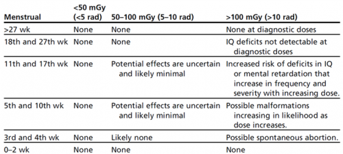
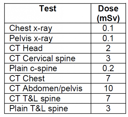
  • Patients with abnormal GCS should undergo CT scanning, consisting of a CT angiogram of the neck and brain with soft tissue images of the neck and cervical spine recons.
  • Based on final diagnoses, the patient can be admitted to an appropriate medical service or mental health. In the very rare case of a spine, airway, or vascular injury, the appropriate service can be consulted.

Reference: A case for less workup in near hanging. J Trauma 81(5):925-930, 2016.

Print Friendly, PDF & Email

Amaze Your Friends! The “Greasy Blood” Sign

Today, I’m writing about a clinical observation that I’ve not seen documented in the doctor books. Maybe it has and I’ve missed it. You be the judge.

I call this particular observation the “greasy blood” sign. You have probably seen it before in your practice as a trauma professional. It is present when you see blood (usually venous) coming from an extremity puncture wound or laceration. What makes it unique is the presence of what looks like drops of oil floating on the surface of the blood.

Here are some learning points about this “greasy blood” sign:

  • What you are actually seeing is fat from bone marrow issuing from an underlying fracture
  • It is most commonly seen in blunt trauma with an open fracture
  • It generally comes from femur or tib/fib fractures, although I’ve seen it a few times from upper extremity fractures
  • If it is associated with a penetrating injury, it is always a gunshot and typically the underlying fracture is very comminuted

Have you seen this sign in your practice? If so, tweet or comment and share any nuances you’ve experienced.

Print Friendly, PDF & Email

Geriatric Week 4: The Medical Orthopaedic Trauma Service

Our population is aging, and falls continue to be a leading cause of injury and morbidity in the elderly. Unfortunately, many elders have significant medical conditions that make them more likely to suffer unfortunate complications from their injuries and the procedures that repair them.

A few hospitals around the world are applying a more multidisciplinary approach than the traditional model. One example is the Medical Orthopaedic Trauma Service (MOTS) at New York-Presbyterian Hospital/Weill Cornell Medical Center. Any elderly patient who has suffered a fracture is seen in the ED by both an emergency physician and a hospitalist from the MOTS team. Once in the hospital, the hospitalist and orthopaedic surgeon try to determine the reason for the fall, assess for risk factors such as osteoporosis, provide comprehensive medical management, provide pain control, and of course, fix the fracture.

This medical center recently published a paper looking at their success with this model. They retrospectively reviewed 306 patients with femur fractures involving the greater trochanter. They looked at complications, length of stay, readmission rate and post-discharge mortality. No change in length of stay was noted, but there were significantly fewer complications, specifically catheter associated urinary tract infections and arrhythmias. The readmission rate was somewhat shorter in the MOTS group, but did not quite achieve significance with regression analysis.

Bottom line: This type of multidisciplinary approach to these fragile patients makes sense. Hospitalists, especially those with geriatric experience, can have a significant impact on the safety and outcomes of these patients. But even beyond this, all trauma professionals need to look for and correct the reasons for the fall, not just fix the bones and send our elders home. This responsibility starts in the field with prehospital providers, and continues with hospital through the entire inpatient stay.

Reference: The medical orthopaedic service (MOTS): an innovative multidisciplinary team model that decreases in-hospital complications in patients with hip fractures. J Orthopaedic Trauma, 26(6):379-383, 2012.

Print Friendly, PDF & Email

Next Week: Trauma In The Elderly

All of next week, I’ll be writing about a topic that is becoming more and more important: geriatric trauma. Our population is aging, and the number of older patients being admitted to trauma centers is exploding.

Here are the topics to be covered:

  • How We Take Care Of Our Elders
  • Thoughts On Geriatric Trauma
  • Elderly Trauma And The Frailty Index
  • The Medical Orthopaedic Trauma Service
  • Falls In The Elderly: The Consequences
  • Effect Of an In-Hospital Falls Prevention Program

And please feel free to leave comments and suggest future topics!

Print Friendly, PDF & Email