Category Archives: General

Cardiac Enzymes And Blunt Cardiac Injury

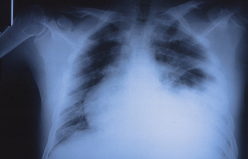
Blunt cardiac injury (BCI), better known as cardiac contusion, has been poorly understood for decades. There has always been confusion about the best way to detect, monitor, and treat this condition. The Eastern Association for the Surgery of Trauma developed a set of practice guidelines in 1998 that provided solid, evidence-based principles to act upon. At that time, CPK was the only cardiac enzyme available. It was useless because it had no correlation with presence or absence of contusion, or even severity of injury.

With the arrival of troponin assays, a new tool for looking at cardiac injury became available. However, initial evaluations indicated that it was also not very reliable when used in the diagnosis of cardiac contusion. For years, I’ve been teaching that these enzymes are useless. However, additional work has been done that does show some limited usefulness.

A prospective study by Collins looked at the usefulness of troponin I (TnI) in patients with cardiac injury. As in previous retrospective studies, this test had a very low positive predictive value. In addition, they determined that a negative EKG alone ruled out blunt cardiac injury. So what good is it?

The new piece of information here is that if the EKG is abnormal and the troponin I is normal, then a cardiac contusion can be ruled out. This changes the overall algorithm by allowing us to eliminate some patients who have abnormal EKGs.

On Monday, I’ll provide my take on a comprehensive algorithm for evaluating and managing cardiac injury. Stay tuned.

Related posts:

Reference: The usefulness of serum troponin levels in evaluating cardiac injury. Am Surg 67:821-826, 2001.

Print Friendly, PDF & Email

Contrast Blush in Children

A contrast blush is occasionally seen on abdominal CT in patients with solid organ injury. This represents active arterial extravasation from the injured organ. In most institutions, this is grounds for call interventional radiology to evaluate and possibly embolize the problem. The image below shows a typical blush.

Splenic contrast blush

This thinking is fairly routine and supported by the literature in adults. However, it cannot be generalized to children!

Children have more elastic tissue in their spleen and tend to do better with nonoperative management than adults. The same holds true for contrast blushes. The vast majority of children will stop bleeding on their own, despite the appearance of a large blush. In fact, if children are taken to angiography, it is commonplace for no extravasation to be seen!

Angiography introduces the risk of local complications in the femoral artery as well as more proximal ones. That, coupled with the fact that embolization is rarely needed, should keep any prudent trauma surgeon from ordering the test. A recently released paper confirms these findings.

The only difficult questions is “when is a child no longer a child?” Is there an age cutoff at which the spleen starts acting like an adult and keeps on bleeding? Unfortunately, we don’t know. I recommend that you use the “eyeball test”, and reserve angiography for kids with contrast extravasation who look like adults (size and body habitus).

Reference: What is the significance of contrast “blush” in pediatric blunt splenic trauma? Davies et al. J Pediatric Surg 2010 May; 45(5):916-20.

Print Friendly, PDF & Email

Pet Peeve: Pain Medication Prescriptions

How much pain medication should you give your patients to take home? The ideal? As much as they need for adequate relief for a reasonable period of time. The reality? Nowhere near enough. 

I see this problem from every level of provider, from interns to senior physicians. I recently underwent surgery on my arm as an outpatient procedure. I was sent home with a prescription for oxycodone 5mg / acetominophen 500mg. It was written to be taken 1-2 tabs q4-6 hours as needed for pain. How many did I get? Twenty!

Now, let’s do the math. If I were to take the maximum 8 per day, this would last me exactly 2.5 days. I’m scheduled to see my provider in 8 days. In the US, this is a Schedule II narcotic, which means my pharmacist needs a paper prescription in his hand to fill it. Phone or fax orders are not acceptable. If I need more before my office visit, I have to get a phone order for a less powerful analgesic, or I have to ask someone to drive to the office and pick up a new paper prescription. For more than 20, I hope. And what if it were a weekend?

Bottom line: DO THE MATH! Give your patients enough medication to get them to their next appointment, commensurate with the amount of pain you expect them to have. For the prescription above 50-60 tabs would have been more appropriate, or a little less (40) if you expected them to taper their dose during the week. Patients with legitimate analgesia needs cannot get addicted in these short time frames for minor to moderate injury.

Related post:

Print Friendly, PDF & Email

Pop Quiz: Delayed Answer!

Well, it took me a month to figure out that one of my pop quiz answers was posted to the wrong area! The link provided with the tweet would have gotten you to it, but those who were following along on tumblr may have been mystified. Here’s the answer again.

Here’s a link to the original post/quiz.

So the question was to guess the exact mechanism of injury given the x-ray below.

The image shows a stomach bubble located in the left chest, indicating a left sided diaphragm rupture. In countries with left sided drivers, this is a classic injury from a t-bone type impact directly on the driver side door. The arm rest is driven into the driver’s flank, or on occasion the driver is partially ejected through the window. The impact raises abdominal pressure abruptly and can push the abdominal contents (typically stomach, followed by spleen) through the weakest area of the diaphragm.

Practical tip: These patients may present with tachypnea and decreased breath sounds on the left side. The usual reflex is to insert a chest tube, which is unneeded and won’t help in this case. What the patient really needs is an NG tube to help their breathing (and an immediate trip to the OR). So if you encounter this clinical combination plus a significant left sided impact (car crash, pedestrian struck), get a chest x-ray first if the patient’s condition will tolerate it. 

And again, hats off to precordialthump for getting it exactly right!

Print Friendly, PDF & Email

Chest Tube Management Protocol – Pediatric

Yesterday I described a protocol for deciding when to remove a chest tube in adults. Today, I’ll go over a variant of this algorithm for children. In general, it’s very similar. The major change is in the volume criterion. In adults, we decided upon an (arbitrary) value of 150cc per three shifts. We chose a time interval of 3 shifts vs 1 day to speed up the process.

Suppose you use the 1 day rule for looking at chest tube output. Typically, this would be evaluated in the morning, and the process of pulling the tube or applying water seal, followed by delayed xrays, could lead to a very late discharge. If the output is checked every shift and the most recent three are summed, the patient could meet criteria later in the day and have the tube pulled in the evening. This would allow for an earlier discharge the following day, shaving 12 hours or more off of the hospital stay. This may not make much of a difference to the hospital (although for busy ones it does), but it’s huge for patient comfort and satisfaction.

Click this image or the link below for a full-size version.

Note that the output criterion has been changed to 2cc/kg over three shifts. This adjusts for the varying sizes of the children that we treat. Otherwise things are basically the same.

Related posts:

Print Friendly, PDF & Email