Thanks for reading!
Michael
Thanks for reading!
Michael
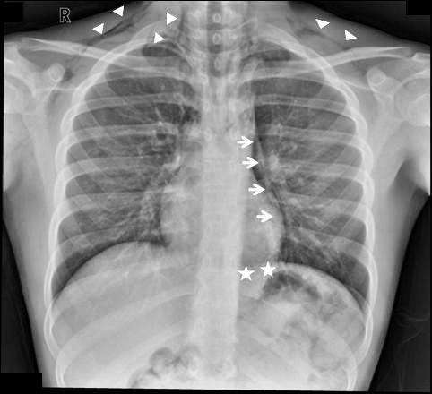
Finding pneumomediastinum on a chest xray or CT scan always gets one’s attention. However, seeing this condition after a simple fall from standing is very simple to evaluate and manage.
There are 3 potential sources of gas in the mediastinum after trauma:
Blunt injury to the esophagus is extremely rare, and probably nonexistent after just falling down. Likewise, a tracheal injury from falling over is unheard of. Both of these injuries are far more common with penetrating trauma.
This leaves the lung and smaller airways within it to consider. They are, by far, the most common sources of pneumomediastinum. The most common pattern is that this injury causes a small pneumothorax, which dissects into the mediastinum over time. On occasion, the leak tracks along the visceral pleura and moves directly to the mediastinum.
Management is simple: a repeat chest xray after 6 hours is needed to show non-progression of any pneumothorax, occult or obvious. This image will usually show that the mediastinal air is diminishing as well. There is no need for the patient to be kept NPO or in bed. Monitor any subjective complaints and if all progresses as expected, they can be discharged after a very brief stay.
Tomorrow: A more interesting (and complicated) case of pneumomediastinum.
The radiologist made me order that (unnecessary) test! I’ve heard this excuse many, many times. Do these phrases look familiar?
Reference: Pitfalls of the vague radiology report. AJR 174(6):1511-1518, 2000.
Our population is aging, and falls continue to be a leading cause of injury and morbidity in the elderly. Unfortunately, many elders have significant medical conditions that make them more likely to suffer unfortunate complications from their injuries and the procedures that repair them.
More and more hospitals around the world are applying a more multidisciplinary approach than the traditional model. One example is the Medical Orthopaedic Trauma Service (MOTS) at New York-Presbyterian Hospital/Weill Cornell Medical Center. Any elderly patient who has suffered a fracture is seen in the ED by both an emergency physician and a hospitalist from the MOTS team. Once in the hospital, the hospitalist and orthopaedic surgeon try to determine the reason for the fall, assess for risk factors such as osteoporosis, provide comprehensive medical management, provide pain control, and of course, fix the fracture.
This medical center published a paper looking at their success with this model. They retrospectively reviewed 306 patients with femur fractures involving the greater trochanter. They looked at complications, length of stay, readmission rate and post-discharge mortality. No change in length of stay was noted, but there were significantly fewer complications, specifically catheter associated urinary tract infections and arrhythmias. The readmission rate was somewhat shorter in the MOTS group, but did not quite achieve significance with regression analysis.
Bottom line: This type of multidisciplinary approach to these fragile patients makes sense. Hospitalists, especially those with geriatric experience, can have a significant impact on the safety and outcomes of these patients. But even beyond this, all trauma professionals need to look for and correct the reasons for the fall, not just fix the bones and send our elders home. This responsibility starts in the field with prehospital providers, and continues with hospital through the entire inpatient stay.
Related post:
Reference: The medical orthopaedic service (MOTS): an innovative multidisciplinary team model that decreases in-hospital complications in patients with hip fractures. J Orthopaedic Trauma 26(6):379-383, 2012.
A bucket handle injury is a type of mesenteric injury of the intestine. The intestine itself separates from the mesentery, leaving a devascularized segment of bowel that looks like the handle on a bucket (get it?).
These injuries can occur after blunt trauma to the abdomen. The force required is rather extreme, so the usual mechanism is motor vehicle crash. In theory, it could occur after a fall from a significant height, and I have seen one case from a wood fragment that was hurled at the abdomen by a malfunctioning lathe.
The mechanics of this injury are related to fixed vs mobile structures in the abdomen. Injuries tend to occur adjacent to areas of the intestine that are fixed, such as the cecum, ligament of Treitz, colonic flexures and rectum. During sudden deceleration, portions of the intestine adjacent to these areas continue to move, pulling on the nearby attachments. This causes the intestine itself to pull off of its mesentery.
Source: personal archive. Not treated at Regions Hospital
The picture above shows multiple bucket handle injuries in one patient. There are 3 injuries in the small bowel, and one involving the entire transverse colon. Note the obviously devascularized segment at the bottom center of the photo.
The terminal ileum is the most common site for bucket handle tears. Proximal jejunum, transverse colon, and sigmoid colon are other possible areas. In my experience, the driver is more likely to sustain an injury to the terminal ileum, and the front passenger to the sigmoid. I think this is due to the location of the buckle, but it’s just my own observation.
Always think about the possibility of this injury in patients with very high speed deceleration. Seat belt marks have a particularly high association with this injury. If your patient has an abnormal exam in the right lower quadrant, or if the CT shows unusual changes there (“dirty” mesenteric fat, thickened bowel wall, extravasation), I recommend a trip to the OR. In these cases, an injury will nearly always be present. And since the affected intestine may take a few days to die and leak, look for an unexplained rise in WBC beginning on hospital day 2.
Related posts: